
Product Name: HSP70
Product Number: AB-NN060-2
| Size: | 50 µg | Price: | 89.00 | |
| $US |
Target Full Name: Heat shock 70 kDa protein 1A
Target Alias: HSP70 1; HSP70 2; HSP70.1; HSP72; HSPA1; HSPA1A; HSPA1B
Product Type Specific: Heat shock/stress protein pan-specific antibody
Antibody Code: NN060-2
Antibody Target Type: Pan-specific
Protein UniProt: P0DMV8
Protein SigNET: P0DMV8
Antibody Type: Monoclonal
Antibody Host Species: Mouse
Antibody Ig Isotype Clone: IgG
Antibody Immunogen Source: Human HSP70
Production Method: Protein G purified
Antibody Modification: Unconjugated. Contact KInexus if you are interest in having the antibody biotinylated or coupled with fluorescent dyes.
Antibody Concentration: 1 mg/ml
Storage Buffer: Phosphate buffered saline pH7.4, 50% glycerol, 0.1% sodium azide
Storage Conditions: For long term storage, keep frozen at -40°C or lower. Stock solution can be kept at +4°C for more than 3 months. Avoid repeated freeze-thaw cycles.
Product Use: Western blotting | Immunohistochemistry | ICC/Immunofluorescence | ELISA | FCM | FACS | IEM | Bl
Antibody Dilution Recommended: WB (1:1000) IHC (1:10,000), ICC/IF (1:1000); optimal dilutions for assays should be determined by the user.
Antibody Potency: Very high potency. Detects a ~70 kDa protein in cell and tissue lysates by Western blotting.
Antibody Species Reactivity: Human | Mouse | Rat | Bovine | C.elegans | Dog | Chicken | Drosophilia | Carp | Guinea pig | Hamster | Monkey | Pig | Rabbit | Sheep
Antibody Positive Control: 1 µg/ml of SMC-100 was sufficient for detection of HSP70 in 20 µg of heat shocked HeLa cell lysate by colorimetric immunoblot analysis using Goat anti-mouse IgG:HRP as the secondary antibody.
Antibody Specificity: High
Antibody Cross Reactivity: Does not cross-react with HSC70 (HSP73).
Related Product 1: HSP70 pan-specific antibody (Cat. No.: AB-NN060-10)
Related Product 2: HSP70/HSC70 pan-specific antibody (Cat. No.: AB-NN060-11)
Related Product 3: HSP70/HSC70 pan-specific antibody (Cat. No.: AB-NN060-12)
Related Product 4: HSP70/HSC70 (pan) pan-specific antibody (Cat. No.: AB-NN060-13)
Related Product 5: HSP70 pan-specific antibody (Cat. No.: AB-NN060-3)
Related Product 6: HSP70 (Crab) pan-specific antibody (Cat. No.: AB-NN060-5)
Related Product 7: HSP70 (P. Falciparum) pan-specific antibody (Cat. No.: AB-NN060-6)
Related Product 8: HSP70 pan-specific antibody (Cat. No.: AB-NN060-7)
Related Product 9: HSP70 pan-specific antibody (Cat. No.: AB-NN060-8)
Related Product 10: HSP70 pan-specific antibody (Cat. No.: AB-NN060-9)
Scientific Background: HSP70 genes encode abundant heat-inducible 70-kDa HSPs (HSP70s). In most eukaryotes HSP70 genes exist as part of a multigene family. They are found in most cellular compartments of eukaryotes including nuclei, mitochondria, chloroplasts, the endoplasmic reticulum and the cytosol, as well as in bacteria. The genes show a high degree of conservation, having at least 5O% identity (2). The N-terminal two thirds of HSP70s are more conserved than the C-terminal third. HSP70 binds ATP with high affinity and possesses a weak ATPase activity which can be stimulated by binding to unfolded proteins and synthetic peptides (3). When HSC70 (constitutively expressed) present in mammalian cells was truncated, ATP binding activity was found to reside in an N-terminal fragment of 44 kDa which lacked peptide binding capacity. Polypeptide binding ability therefore resided within the C-terminal half (4). The structure of this ATP binding domain displays multiple features of nucleotide binding proteins (5).
All HSP70s, regardless of location, bind proteins, particularly unfolded ones. The molecular chaperones of the HSP70 family recognize and bind to nascent polypeptide chains as well as partially folded intermediates of proteins preventing their aggregation and misfolding. The binding of ATP triggers a critical conformational change leading to the release of the bound substrate protein (6). The universal ability of HSP70s to undergo cycles of binding to and release from hydrophobic stretches of partially unfolded proteins determines their role in a great variety of vital intracellular functions such as protein synthesis, protein folding and oligomerization and protein transport. Looking for more information on HSP70? Visit our new HSP70 Scientific Resource Guide at http://www.HSP70.com.
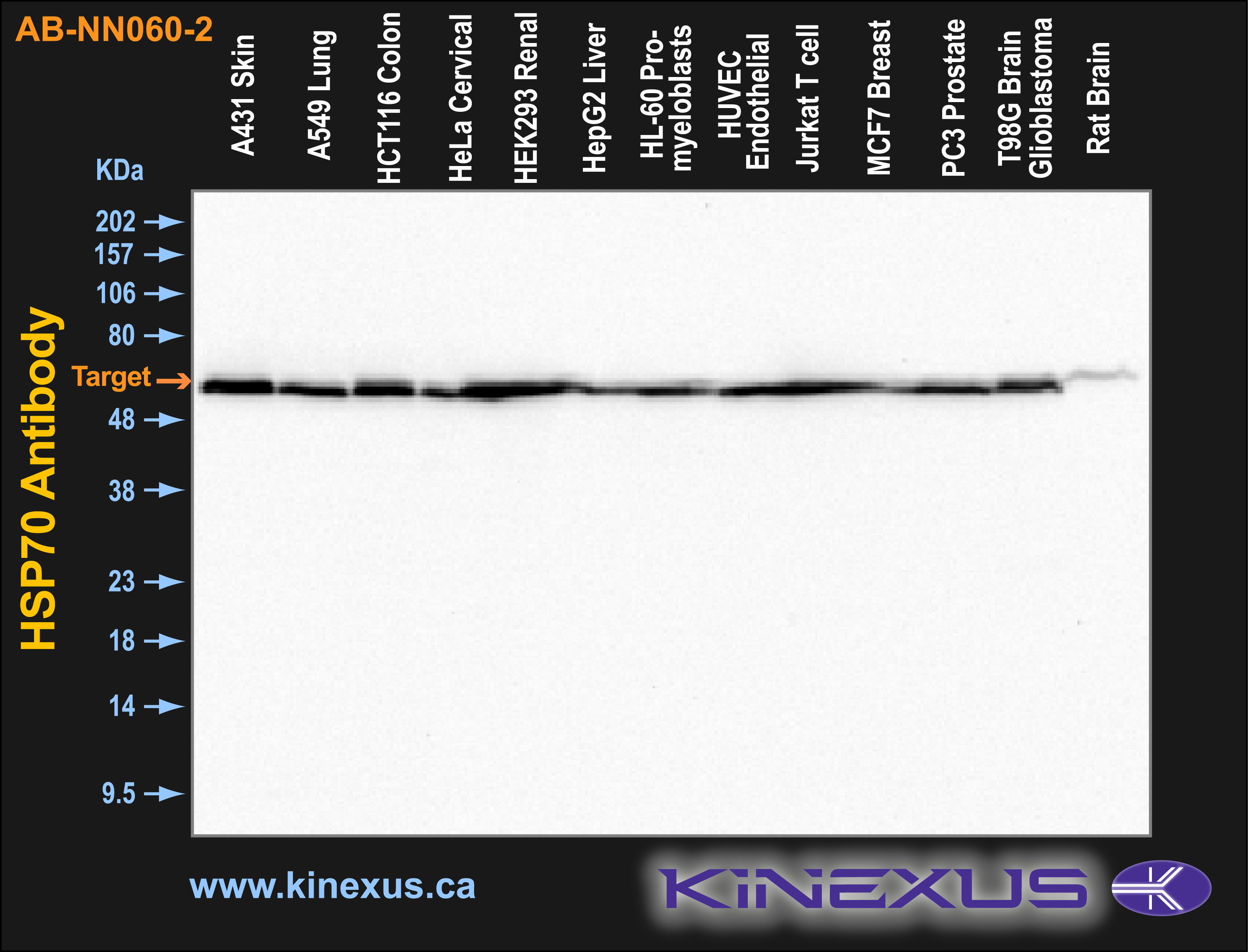
Figure 1. Immunoblotting of various cell lines with AB-NN060-2 antibody at 1 µg/mL final concentration. The target protein Hsp70 is indicated. Each lane was loaded with 15 µg of cell lysate protein. The max signal count was 29429.
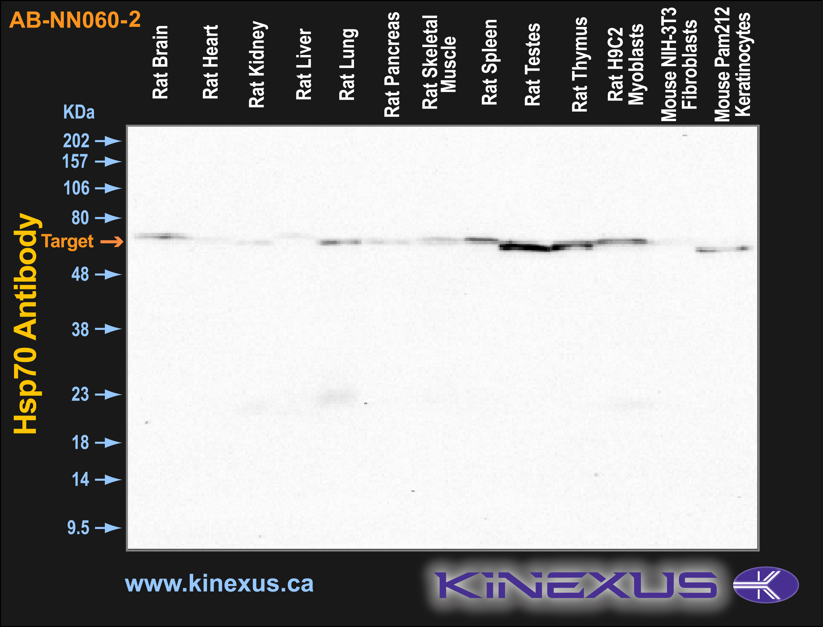
Figure 2. Immunoblotting of various tissue lines with AB-NN060-2 antibody at 1 µg/mL final concentration. The target protein Hsp70 is indicated. Each lane was loaded with 15 µg of tissue lysate protein. The max signal count was 39395.
References
[1] Welch W.J. and Suhan J.P. (1986) J Cell Biol. 103: 2035-2050.
[2] Boorstein W. R., Ziegelhoffer T. & Craig E. A. (1993) J.Mol. Evol. 38(1): 1-17.
[3] Rothman J. (1989) Cell 59: 591-601.
[4] DeLuca-Flaherty et al. (1990) Cell 62: 875-887.
[5] Bork P., Sander C. & Valencia A. (1992) Proc. Nut1 Acad. Sci. USA 89: 7290-7294.
[6] Fink A.L. (1999) Physiol. Rev. 79: 425-449.
[7] Galan A. et al. (2000) J. Biol. Chem. 275: 11418-11424.
[8] Kondo T. et al. (2000) J. Biol. Chem. 275: 8872-8879.
[9] Misaki T. et al. (1994) Clin. Exp. Immun. 98: 234-239.
[10] Pockley A.G. et al. (1998) Immunol. Invest. 27: 367-377.
[11] Moon I.S. et al. (2001) Cereb Cortex 11(3): 238-248.
[12] Dressel et al. (2000) J. Immunol. 164: 2362-2371. Verma A.K. et al. (2007) Fish and Shellfish Immunology. 22(5): 547-555. Banduseela V.C. et al. (2009) Physiol Genomics. 39(3): 141-159.

© Kinexus Bioinformatics Corporation 2017