
Product Name: Ubiquitin
Product Number: AB-NN154-3
| Size: | 50 µg | Price: | 89.00 | |
| $US |
Target Full Name: Ubiquitin
Target Alias: Polyubiquitin B; RPS27A; UBA52; UBB; UBC; ubiquitin B
Product Type Specific: Ubiquitin pan-specific antibody
Antibody Code: NN154-3
Antibody Target Type: Pan-specific
Protein UniProt: P0CG47
Protein SigNET: P0CG47
Antibody Type: Monoclonal
Antibody Host Species: Mouse
Antibody Ig Isotype Clone: IgG2a Kappa
Antibody Immunogen Source: Native bovine ubiquitin, conjugated to KLH
Production Method: Protein G purified
Antibody Modification: Unconjugated. Contact KInexus if you are interest in having the antibody biotinylated or coupled with fluorescent dyes.
Antibody Concentration: 1 mg/ml
Storage Buffer: Phosphate buffered saline pH7.4, 50% glycerol, 0.09% sodium azide
Storage Conditions: For long term storage, keep frozen at -40°C or lower. Stock solution can be kept at +4°C for more than 3 months. Avoid repeated freeze-thaw cycles.
Product Use: Western blotting | ICC/Immunofluorescence | ELISA
Antibody Dilution Recommended: WB (1:1000), ICC/IF (1:100); optimal dilutions for assays should be determined by the user.
Antibody Potency: High potency. Detects a ~10 kDa protein in cell and tissue lysates by Western blotting.
Antibody Species Reactivity: Human | Mouse | Rat | Bovine
Antibody Positive Control: 1 µg/ml of SMC-160 was sufficient for detection of ubiquitin in 10 µg of Heal Lysate by colorimetric immunoblot analysis using Goat anti-mouse IgG:HRP as the secondary antibody.
Antibody Specificity: Very high
Related Product 1: Ubiquitin pan-specific antibody (Cat. No.: AB-NN154-2)
Scientific Background: Ubiquitin is a small protein that occurs in all eukaryotic cells. The ubiquitin protein itself consists of 76 amino acids and has a molecular mass of about 8.5kDa. Key features include its C-terminal tail and the 7 Lys residues. It is highly conserved among eukaryotic species: Human and yeast ubiquitin share 96% sequence identity (1). The main function of Ubiquitin is to clear abnormal, foreign and improperly folded proteins by targeting them for degradation by the 26S proteosome (2). Ubiquitination represents an essential cellular process affected by a multi-enzyme cascade involving classes of enzymes known as ubiquitin-activating enzymes (E1s), ubiquitin-conjugating enzymes (E2s or Ubcs) and ubiquitin-protein ligases (E3s). Ubiquitin is activated in a two-step reaction by an E1 ubiquitin-activating enzyme in a process requiring ATP as an energy source. The initial step involves production of an ubiquitin-adenylate intermediate. The second step transfers ubiquitin to the E1 active site cysteine residue, with release of AMP. This step results in a thioester linkage between the C-terminal carboxyl group of ubiquitin and the E1 cysteine sulfhydryl group. The third step is a transfer of ubiquitin from E1 to the active site cysteine of a ubiquitin-conjugating enzyme E2 via a trans(thio)esterification reaction. And the final step of the ubiquitylation cascade creates an isopeptide bond between a lysine of the target protein and the C-terminal glycine of ubiquitin. In general, this step requires the activity of one of the hundreds of E3 ubiquitin-protein ligases (often termed simply ubiquitin ligase). E3 enzymes function as the substrate recognition modules of the system and are capable of interaction with both E2 and substrate(2, 3). Ubiquitination also participates in the internalization and degradation of plasma membrane proteins such as some of the TCR subunits while still ER-membrane associated (4). Ubiquitin also plays a role in regulating signal transduction cascades through the elimination inhibitory proteins, such as IκBα and p27 (5).
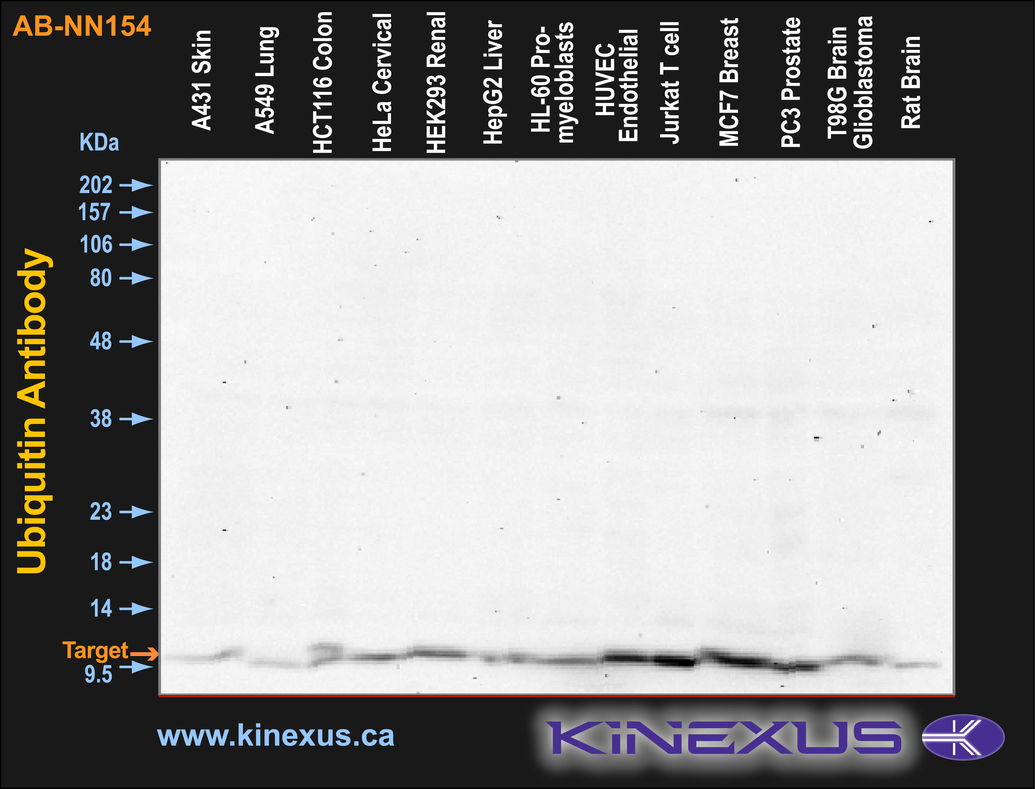
Figure 1. Immunoblotting of various cell lines with AB-NN154 antibody at 25 X dilution. The target protein Ubiquitin is indicated. Each lane was loaded with 15 µg of cell lysate protein. The max signal count was 5789.
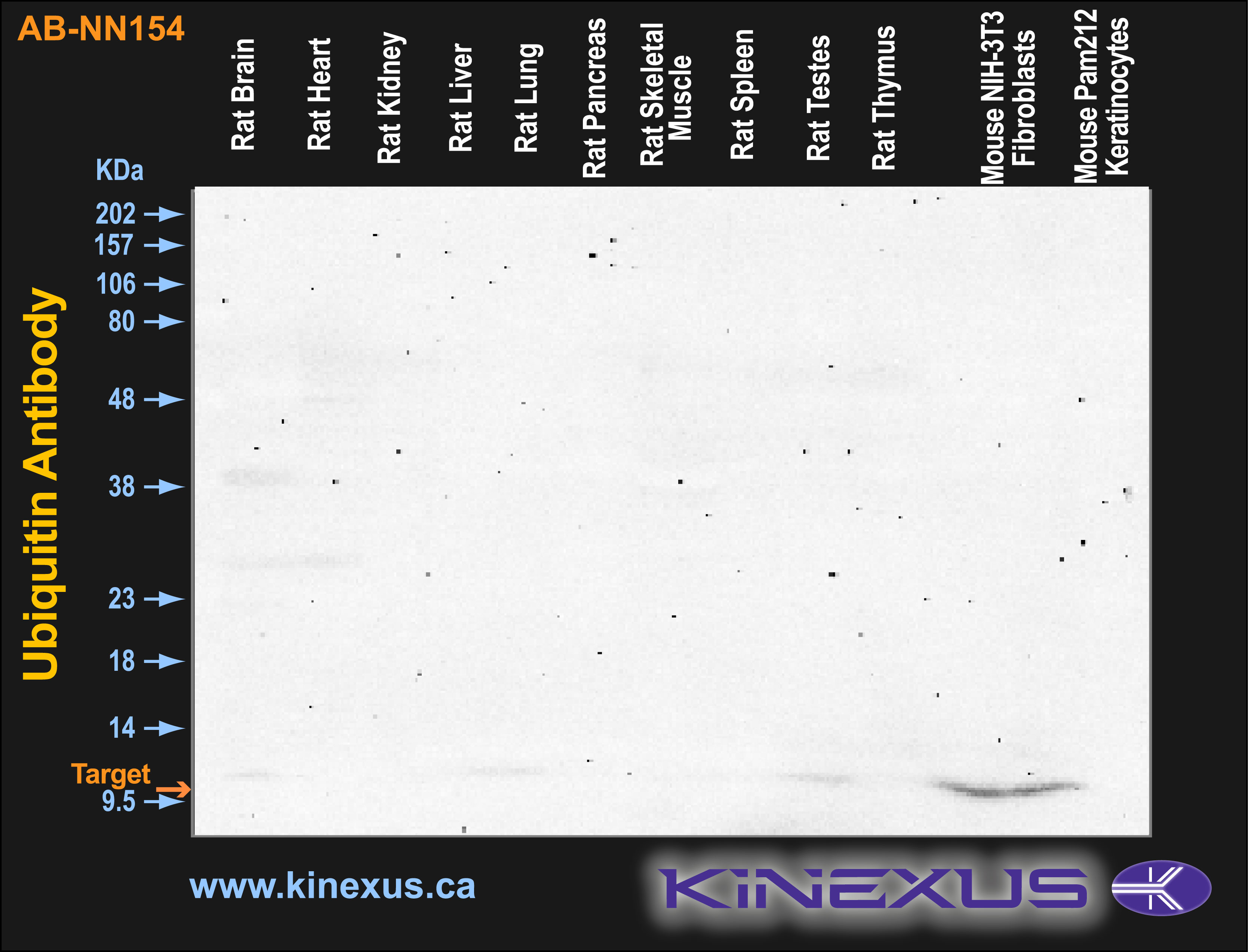
Figure 2. Immunoblotting of various tissue lines with AB-NN154 antibody at 25 X dilution. The target protein Ubiquitin is indicated. Each lane was loaded with 15 µg of tissue lysate protein. The max signal count was 4147.
References
[1] Wilkinson K.D. (1995) Annu. Rev. Nutr. 15:161-189.
[2] Bonifacino J.S. et al. (1998) Annu Rev Cell Dev Biol. 14: 19-57.
[3] Boston Biochem: "Ubiquitin Proteasome Pathway Overview” http://www.bostonbiochem.com/upp.php
[4] Yang M. et al. (1998) J Exp Med. 187: 1835-1846.
[5] Chen Z.J. et al. (1996) Cell 84: 853-862.

© Kinexus Bioinformatics Corporation 2017