
Product Name: Cot-pS334
Product Number: AB-PK585
| Size: | 25 µg | Price: | 89.00 | |
| $US |
Target Full Name: Osaka thyroid oncogene protein-serine kinase (Tpl2); Mitogen-activated protein kinase kinase kinase 8
Target Alias: FLJ10486; M3K8; MAP3K8; Tpl2; TPL2; TPL-2; tumour progression locus 2; Cot; ESTF; EST; Tpl-2; c-Cot; ENSG00000107968
Product Type Specific: Protein kinase phosphosite-specific antibody
Antibody Code: PK585
Antibody Target Type: Phosphosite-specific
Antibody Phosphosite: S334
Protein UniProt: P41279
Protein SigNET: P41279
Antibody Type: Polyclonal
Antibody Host Species: Rabbit
Antibody Immunogen Source: Human Cot (MAP3K8, TPL2) sequence peptide Cat. No.: PE-04AKB90
Antibody Immunogen Sequence: YPR(pS)AYP(bA)C
Antibody Immunogen Description: Corresponds to amino acid residues Y331 to P337; In the kinase catalytic subdomain X region.
Antibody Immunogen Sequence: YPR(pS)AYP(bA)C
Antibody Immunogen Description: Corresponds to amino acid residues Y331 to P337; In the kinase catalytic subdomain X region.
Production Method: The immunizing peptide was produced by solid phase synthesis on a multipep peptide synthesizer and purified by reverse-phase hplc chromatography. Purity was assessed by analytical hplc and the amino acid sequence confirmed by mass spectrometry analysis. This peptide was coupled to KLH prior to immunization into rabbits. New Zealand White rabbits were subcutaneously injected with KLH-coupled immunizing peptide every 4 weeks for 4 months. The sera from these animals was applied onto an agarose column to which the immunogen peptide was thio-linked. Antibody was eluted from the column with 0.1 M glycine, pH 2.5. Subsequently, the antibody solution was neutralized to pH 7.0 with saturated Tris.This antibody was also subject to negative purification over phosphotyrosine-agarose.
Antibody Modification: Unconjugated. Contact KInexus if you are interest in having the antibody biotinylated or coupled with fluorescent dyes.
Antibody Modification: Unconjugated. Contact KInexus if you are interest in having the antibody biotinylated or coupled with fluorescent dyes.
Antibody Concentration: 1 mg/ml
Storage Buffer: Phosphate buffered saline pH 7.4, 0.05% Thimerasol
Storage Conditions: For long term storage, keep frozen at -40°C or lower. Stock solution can be kept at +4°C for more than 3 months. Avoid repeated freeze-thaw cycles.
Product Use: Western blotting | Antibody microarray
Antibody Dilution Recommended: 2 µg/ml for immunoblotting
Antibody Potency: Medium-weak immunoreactivity of a target-sized protein by Western blotting in A431 cells. Very strong immunoreactivity with immunogen peptide on dot blots.
Antibody Species Reactivity: Human
Antibody Positive Control: The observed molecular mass of the processed target protein on SDS-PAGE gels is reported to be around 52-57 kDa.
Antibody Specificity: Very high
Antibody Cross Reactivity: No significant cross-reactive proteins detected in A431 cells and in sea star oocytes.
Related Product 1: Cot-pS334 blocking peptide
Related Product 2: Cot pan-specific antibody (Cat. No.: AB-NK042)
Related Product 3: Cot-CT (Cot-2) pan-specific antibody (Cat. No.: AB-NK042-2)
Related Product 4: Cot (Cot-1) pan-specific antibody (Cat. No.: AB-NK042-1)
Scientific Background: Cot (TPL2, MAP3K8) is a protein-serine/threonine kinase of the STE group and STE-Unique family. It is essential in the TLR4 (recognizing LPS) MAPK/ERK-mediated response in the production of pro-inflammatory cytokines. Catalytic activity of Cot is lost with the K167R, or D270A mutations. IL-1 stimulated activity is decreased with S62A. This mutation will also inhibit IL-1 stimulated phosphorylation of T290 along with MEK phosphorylation when associated with A400. Full inhibition of IL-1 induced MEK phosphorylation and partial inhibition of autophosphorylation can occur with a T290A mutation. Partial inhibition of MEK phosphorylation and autophosphorylation has occurred with a T290A or T290E mutation. Partial impairment of MEK phosphorylation induced by IL-1 has been observed with a S400A mutation. When Cot is associated with A62, a complete loss of T290 and MEK phosphorylation occurs with a S400A mutation.
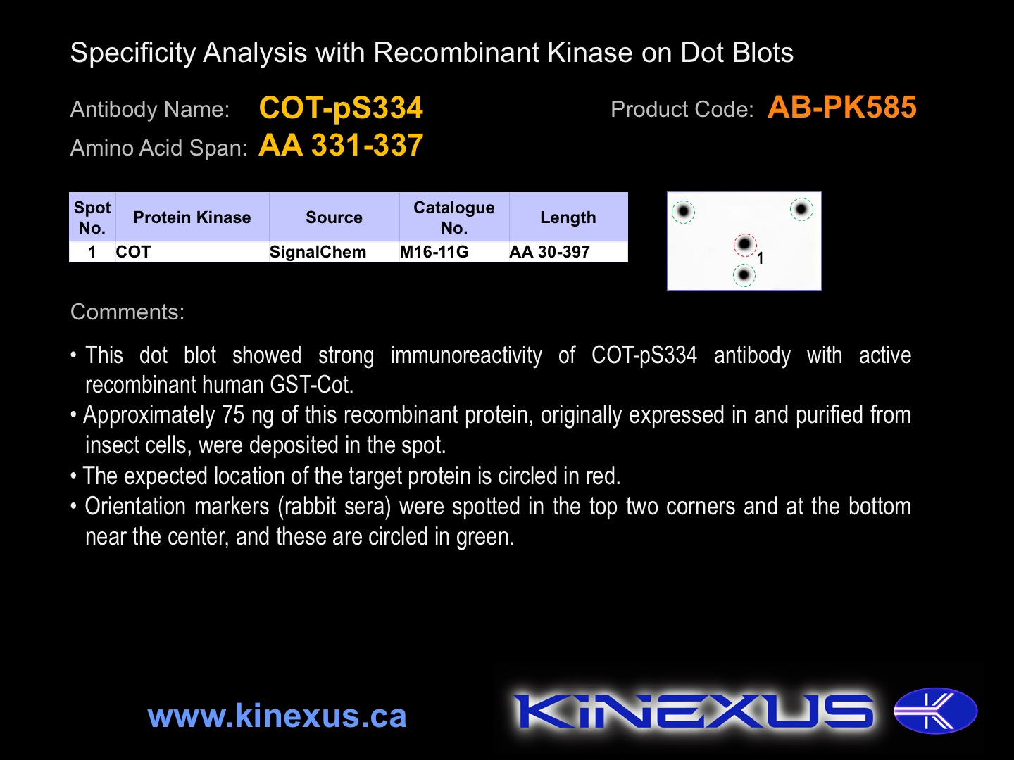
Figure 1. Dot blotting COT-pS334 antibody with recombinant purified proteins.
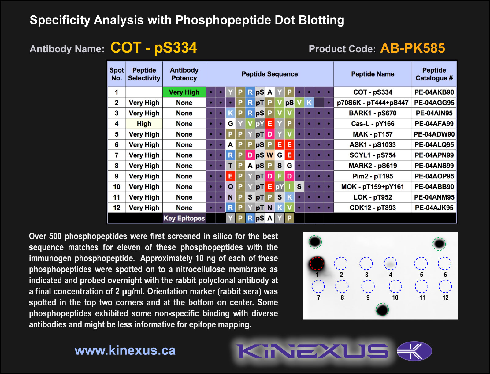
Figure 2. Epitope mapping of COT-pS334 antibody with similar phosphopeptides on dot blots.
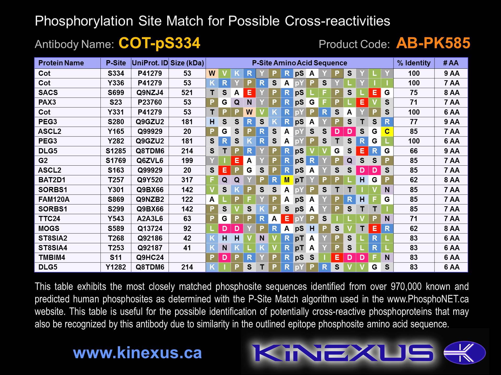
Figure 3. Identification of phosphosites related to COT-pS334.

© Kinexus Bioinformatics Corporation 2017