
Product Name: DYRK1A-pY321
Product Number: AB-PK597
| Size: | 25 µg | Price: | 89.00 | |
| $US |
Target Full Name: Dual specificity tyrosine-phosphorylation-regulated kinase 1A
Target Alias: DYR1A; DYRK; MNB; MNBH; HP86; DYRK1; CCDS13653.1; ENSG00000157540
Product Type Specific: Protein kinase phosphosite-specific antibody
Antibody Code: PK597
Antibody Target Type: Phosphosite-specific
Antibody Phosphosite: Y321
Protein UniProt: Q13627
Protein SigNET: Q13627
Antibody Type: Polyclonal
Antibody Host Species: Rabbit
Antibody Immunogen Source: Human DYRK1A sequence peptide Cat. No.: PE-04AET99
Antibody Immunogen Sequence: IYQ(pY)IQS(bA)C
Antibody Immunogen Description: Corresponds to amino acid residues I318 to S324; In the protein kinase catalytic domain activation T loop region between subdomains VII and VIII.
Antibody Immunogen Description: Corresponds to amino acid residues I318 to S324; In the protein kinase catalytic domain activation T loop region between subdomains VII and VIII.
Production Method: The immunizing peptide was produced by solid phase synthesis on a multipep peptide synthesizer and purified by reverse-phase hplc chromatography. Purity was assessed by analytical hplc and the amino acid sequence confirmed by mass spectrometry analysis. This peptide was coupled to KLH prior to immunization into rabbits. New Zealand White rabbits were subcutaneously injected with KLH-coupled immunizing peptide every 4 weeks for 4 months. The sera from these animals was applied onto an agarose column to which the immunogen peptide was thio-linked. Antibody was eluted from the column with 0.1 M glycine, pH 2.5. Subsequently, the antibody solution was neutralized to pH 7.0 with saturated Tris.This antibody was also subject to negative purification over phosphotyrosine-agarose.
Antibody Modification: Unconjugated. Contact KInexus if you are interest in having the antibody biotinylated or coupled with fluorescent dyes.
Antibody Modification: Unconjugated. Contact KInexus if you are interest in having the antibody biotinylated or coupled with fluorescent dyes.
Antibody Concentration: 0.3 mg/ml
Storage Buffer: Phosphate buffered saline pH 7.4, 0.05% Thimerasol
Storage Conditions: For long term storage, keep frozen at -40°C or lower. Stock solution can be kept at +4°C for more than 3 months. Avoid repeated freeze-thaw cycles.
Product Use: Western blotting | Antibody microarray
Antibody Dilution Recommended: 2 µg/ml for immunoblotting
Antibody Potency: Very weak immunoreactivity of a target-sized protein by Western blotting in A431 and HEK-293 cells. Medium immunoreactivity with immunogen peptide on dot blots.
Antibody Species Reactivity: Human
Antibody Positive Control: The observed molecular mass of the processed target protein on SDS-PAGE gels is reported to be around 80-90 kDa.
Antibody Specificity: Medium-High
Antibody Cross Reactivity: At high concentrations, this antibody detects multiple proteins (~85, ~37 and ~28 kDa) in A431 cells.
Related Product 1: DYRK1A-pY321 blocking peptide
Related Product 2: DYRKSubtide - DYRK1A protein kinase substrate peptide
Antibody Specificity: Medium-High
Antibody Cross Reactivity: At high concentrations, this antibody detects multiple proteins (~85, ~37 and ~28 kDa) in A431 cells.
Related Product 1: DYRK1A-pY321 blocking peptide
Related Product 2: DYRKSubtide - DYRK1A protein kinase substrate peptide
Scientific Background: DYRK1A is a protein-serine/threonine kinase of the CMGC group and DYRK family. It is a dual-specificity protein kinase that can phosphorylate tyrosine, serine, and threonine residues, as well as the proteins CRY2, FOXO1, SRSF6 and SIRT1. Phosphorylation at Y321 increases phosphotransferase activity. Phosphorylation at S529 also increases phosphotransferase activity and association with 14-3-3-beta. It is inhibited by RANBP9. It may play a role in cell proliferation pathways such as during brain development. DYRK1A has been found to prolong ERK activation via interactions with RAS, BRAF, and MEK1 and the formation of a RAS/BRAF/MEK1 complex. Overexpression in pheochromocytoma cells potentiated their neuronal differentiation in response to NGF through a RAS/MAPK signalling pathway. DYRK1A may also interact with viral oncoproteins such as adenovirus E1A and HPV-E7. Its role in cancer is unclear: inhibition of caspase-9 by DYRK1A protects mitotic cells from apoptosis, but overexpression attentuated calcineurin signalling, which leads to decreased angiogenesis and tumour growth. Active DYRK1A can induce megakaryocytic tumours (derived from bone marrow cells responsible for producing red blood cells).
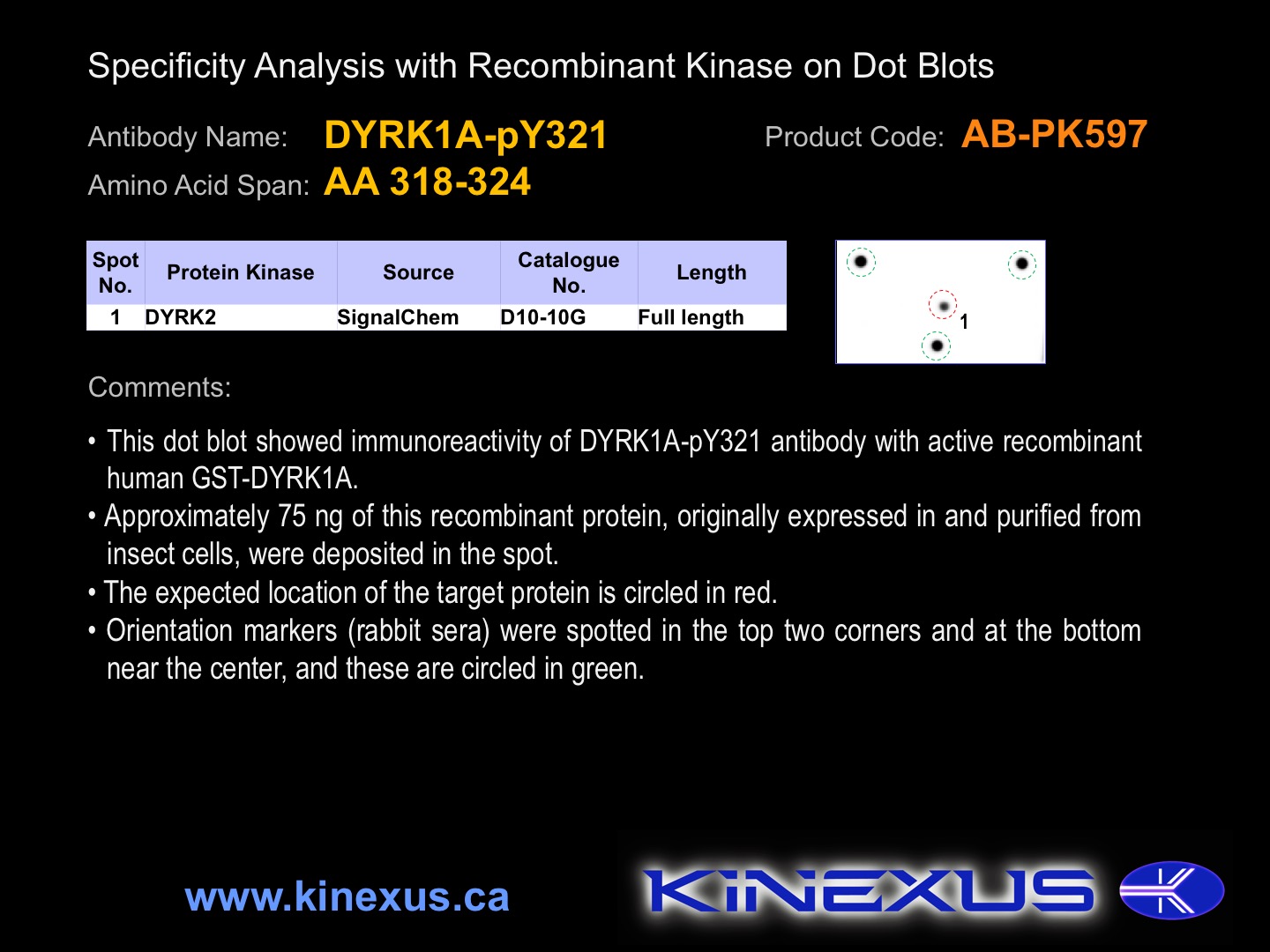
Figure 1. Dot blotting DYRK1A-pY321 antibody with recombinant purified proteins.
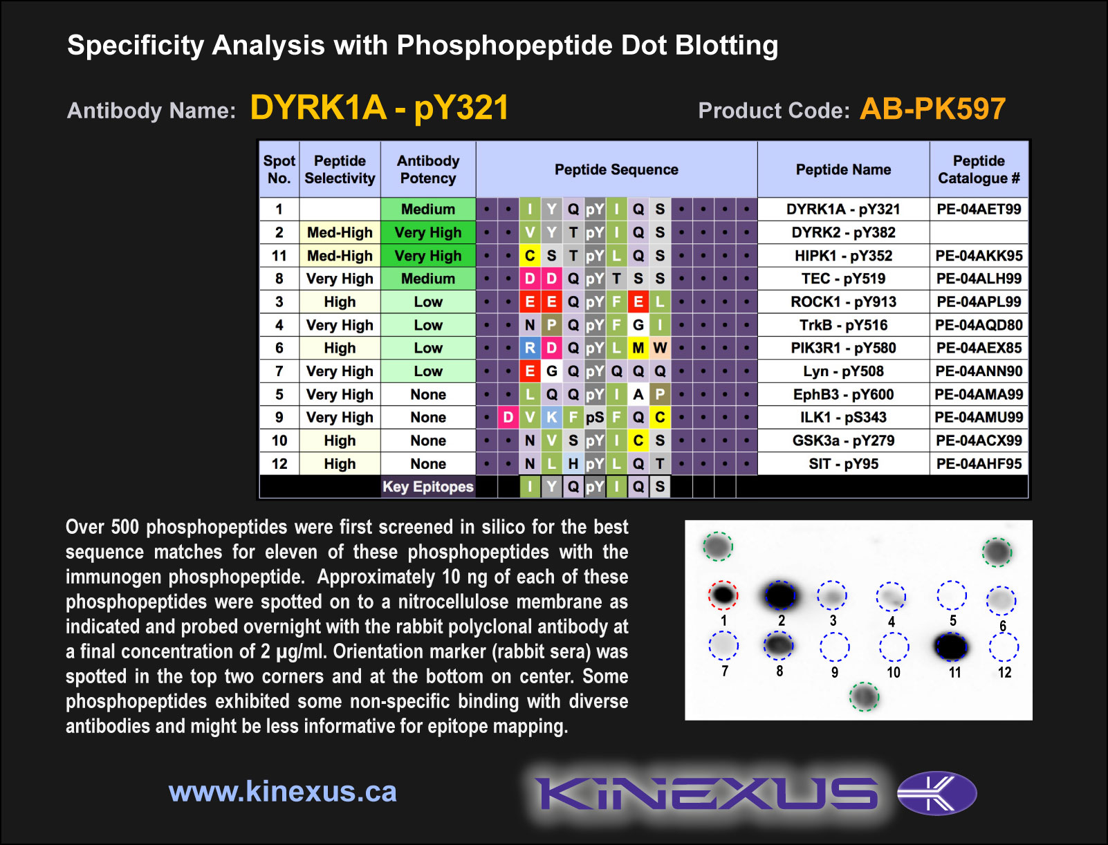
Figure 2. Epitope mapping of DYRK1A-pY321 antibody with similar phosphopeptides on dot blots.
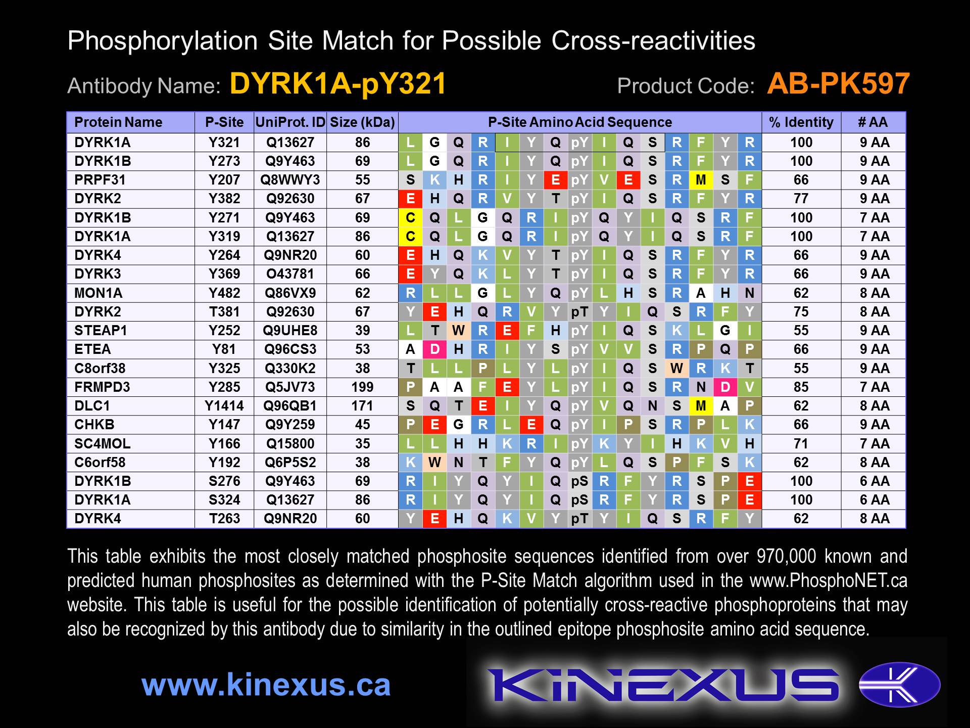
Figure 3. Identification of phosphosites related to DYRK1A-pY321.

© Kinexus Bioinformatics Corporation 2017