
Product Name: Fes-pY713+pS716
Product Number: AB-PK633
| Size: | 25 µg | Price: | 89.00 | |
| $US |
Target Full Name: Fes/Fps protein-tyrosine kinase
Target Alias: C-Fes; C-fes/fps protein; Feline sarcoma oncogene; Fes/FPS; FPS; Kinase Fes; Oncogene Fes, feline sarcoma virus; CCDS10365.1; ENSG00000182511
Product Type Specific: Protein kinase phosphosite-specific antibody
Antibody Code: PK633
Antibody Target Type: Phosphosite-specific
Antibody Phosphosite: Y713+S716
Protein UniProt: P07332
Protein SigNET: P07332
Antibody Type: Polyclonal
Antibody Host Species: Rabbit
Antibody Immunogen Source: Human Fes sequence peptide Cat. No.: PE-04AFT90
Antibody Immunogen Sequence: GV(pY)AA(pS)GGSL(bA)C
Antibody Immunogen Description: Corresponds to amino acid residues G711 to L720; In the protein kinase catalytic domain activation T loop region between subdomains VII and VIII.
Antibody Immunogen Sequence: GV(pY)AA(pS)GGSL(bA)C
Antibody Immunogen Description: Corresponds to amino acid residues G711 to L720; In the protein kinase catalytic domain activation T loop region between subdomains VII and VIII.
Production Method: The immunizing peptide was produced by solid phase synthesis on a multipep peptide synthesizer and purified by reverse-phase hplc chromatography. Purity was assessed by analytical hplc and the amino acid sequence confirmed by mass spectrometry analysis. This peptide was coupled to KLH prior to immunization into rabbits. New Zealand White rabbits were subcutaneously injected with KLH-coupled immunizing peptide every 4 weeks for 4 months. The sera from these animals was applied onto an agarose column to which the immunogen peptide was thio-linked. Antibody was eluted from the column with 0.1 M glycine, pH 2.5. Subsequently, the antibody solution was neutralized to pH 7.0 with saturated Tris.This antibody was also subject to negative purification over phosphotyrosine-agarose.
Antibody Modification: Unconjugated. Contact KInexus if you are interest in having the antibody biotinylated or coupled with fluorescent dyes.
Antibody Modification: Unconjugated. Contact KInexus if you are interest in having the antibody biotinylated or coupled with fluorescent dyes.
Antibody Concentration: 1 mg/ml
Storage Buffer: Phosphate buffered saline pH 7.4, 0.05% Thimerasol
Storage Conditions: For long term storage, keep frozen at -40°C or lower. Stock solution can be kept at +4°C for more than 3 months. Avoid repeated freeze-thaw cycles.
Product Use: Western blotting | Antibody microarray
Antibody Dilution Recommended: 2 µg/ml for immunoblotting
Antibody Potency: Medium immunoreactivity of a target-sized protein by Western blotting in HEK-293 cells, which is forskolin-induced and in EGF-treated HeLa cells.
Antibody Species Reactivity: Human
Antibody Positive Control: The observed molecular mass of the processed target protein on SDS-PAGE gels is reported to be around 93-100 kDa.
Antibody Specificity: High
Antibody Cross Reactivity: No significant cross-reactive proteins detected in MCF7 cells, but a medium immunoreactive 150 kDa protein was detected in HEK-293 and HeLa cells.
Related Product 1: Fes-pY713+pS716 blocking peptide
Related Product 2: Fes-pY713 phosphosite-specific antibody (Cat. No.: AB-PK632)
Related Product 3: FesSubtide - Fes protein kinase substrate peptide
Scientific Background: Fes is a protein-tyrosine kinase of the TK group and Fer family. It functions downstream of membrane bound receptors and is involved in the regulation of the actin cytoskeleton, microtubule assembly, cell adhesion, and cell spreading. Additionally, Fes plays a role in the degranulation of mast cells and acts downstream of the FCER1 receptor and the mast/stem cell growth factor receptor Kit. In addition, Fes is involved in the regulation of cell differentiation as well as in the promotion of neurite growth in response to nerve growth factor (NGF) signalling. Known targets of the Fes protein include BCR, HCLS1/HS1, PECAM1, STAT3, and TRIM28. Phosphorylation of Y713 increases phosphotransferase activity and interaction with Fes. Fes has been demonstrated to be an oncoprotein in several human cancer types, potentially due to abnormal activation of the kinase catalytic function. It was originally identified as an oncoprotein from avian retroviral (Fps in Fujinami sarcoma virus) and cat retroviral (Fes in feline sarcoma virus) studies. Interestingly, Fes has also been shown to act as a tumour-suppressor protein in some human cancer types. For example, reduced or absent expression of the Fes protein is observed in colon cancer specimens and the ectopic expression of Fes in colon cancer cell lines results in the suppression of anchorage-independent cell growth, indicating a tumour suppressing role for the Fes protein. In contrast, Fes expression is significantly elevated in prostate cancer specimens and is thought to actively promote cell growth in renal carcinoma cell lines. In animal studies, mice lacking Fes expression displayed deficits in hematopoiesis and had significantly reduced numbers of circulating mature myeloid cells and immature myeloid precursors, indicating a oncogenic role for the Fes protein in this cancer.
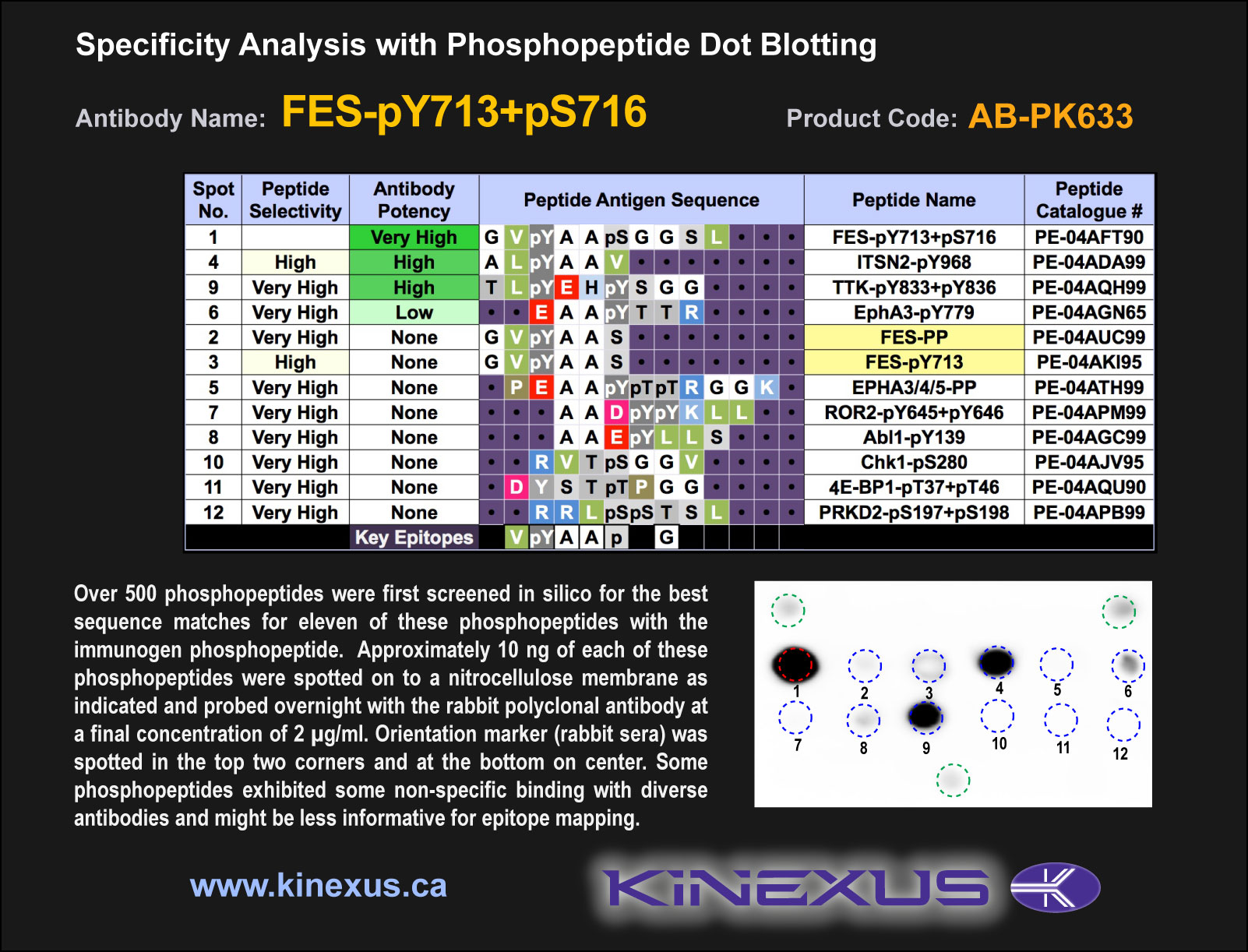
Figure 1. Epitope mapping of FES-pY713+pS716 antibody with similar phosphopeptides on dot blots.
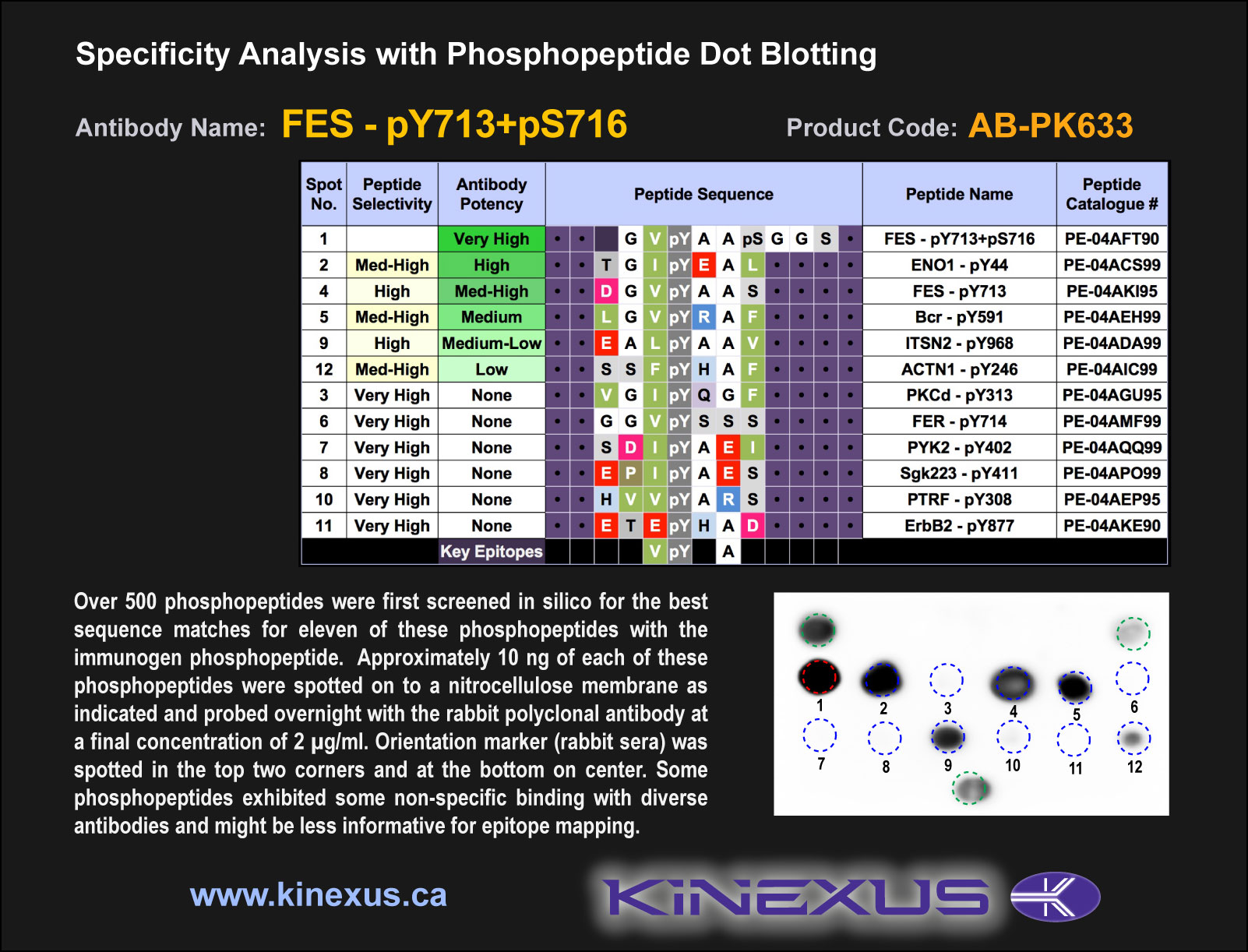
Figure 2. Epitope mapping of FES-pY713+pS716 antibody with similar phosphopeptides on dot blots.
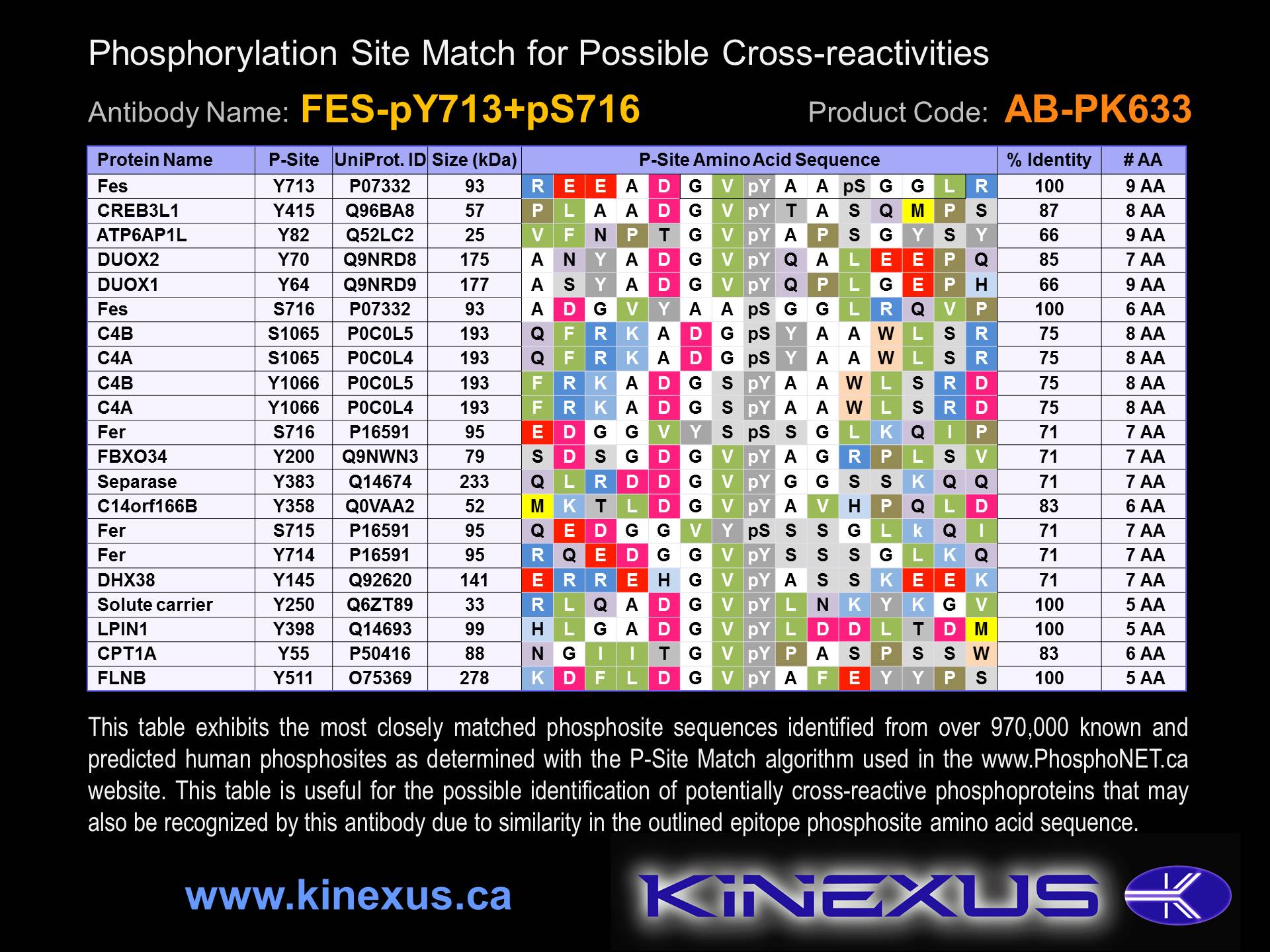
Figure 3. Identification of phosphosites related to FES-pY713+pS716.

© Kinexus Bioinformatics Corporation 2017