
Product Name: GCK-pS170
Product Number: AB-PK646
| Size: | 25 µg | Price: | 89.00 | |
| $US |
Target Full Name: Germinal centre protein-serine kinase; Mitogen-activated protein kinase kinase kinase kinase 2
Target Alias: B lymphocyte serine/threonine-protein kinase; BL44; GCK; Germinal center kinase; Kinase GCK; M4K2; MAP4K2; MAPK/ERK kinase kinase kinase 2; RAB8IP; CCDS8082.1; Q12851; ENSG00000168067
Product Type Specific: Protein kinase phosphosite-specific antibody
Antibody Code: PK646
Antibody Target Type: Phosphosite-specific
Antibody Phosphosite: S170
Protein UniProt: Q12851
Protein SigNET: Q12851
Antibody Type: Polyclonal
Antibody Host Species: Rabbit
Antibody Immunogen Source: Human GCK (MAP4K2) sequence peptide Cat. No.: PE-04AMQ99
Antibody Immunogen Sequence: KRR(pS)FIG(bA)C
Antibody Immunogen Description: Corresponds to amino acid residues K167 to G173; In the protein kinase catalytic domain activation T loop region between subdomains VII and VIII.
Antibody Immunogen Sequence: KRR(pS)FIG(bA)C
Antibody Immunogen Description: Corresponds to amino acid residues K167 to G173; In the protein kinase catalytic domain activation T loop region between subdomains VII and VIII.
Production Method: The immunizing peptide was produced by solid phase synthesis on a multipep peptide synthesizer and purified by reverse-phase hplc chromatography. Purity was assessed by analytical hplc and the amino acid sequence confirmed by mass spectrometry analysis. This peptide was coupled to KLH prior to immunization into rabbits. New Zealand White rabbits were subcutaneously injected with KLH-coupled immunizing peptide every 4 weeks for 4 months. The sera from these animals was applied onto an agarose column to which the immunogen peptide was thio-linked. Antibody was eluted from the column with 0.1 M glycine, pH 2.5. Subsequently, the antibody solution was neutralized to pH 7.0 with saturated Tris.This antibody was also subject to negative purification over phosphotyrosine-agarose.
Antibody Modification: Unconjugated. Contact KInexus if you are interest in having the antibody biotinylated or coupled with fluorescent dyes.
Antibody Modification: Unconjugated. Contact KInexus if you are interest in having the antibody biotinylated or coupled with fluorescent dyes.
Antibody Concentration: 1 mg/ml
Storage Buffer: Phosphate buffered saline pH 7.4, 0.05% Thimerasol
Storage Conditions: For long term storage, keep frozen at -40°C or lower. Stock solution can be kept at +4°C for more than 3 months. Avoid repeated freeze-thaw cycles.
Product Use: Western blotting | Antibody microarray
Antibody Dilution Recommended: 2 µg/ml for immunoblotting
Antibody Potency: Very strong immunoreactivity with immunogen peptide on dot blots.
Antibody Species Reactivity: Human
Antibody Positive Control: The observed molecular mass of the processed target protein on SDS-PAGE gels is reported to be around 85-95 kDa.
Antibody Specificity: Very high
Antibody Cross Reactivity: No significant cross-reactive proteins detected in Jurkat and MCF7 cells.
Related Product 1: GCK-pS170 blocking peptide
Antibody Cross Reactivity: No significant cross-reactive proteins detected in Jurkat and MCF7 cells.
Related Product 1: GCK-pS170 blocking peptide
Scientific Background: GCK (MAP4K2) is a protein-serine/threonine kinase of the STE group and STE20 family. It participates in the regulation of the MAPK signalling pathway. It acts as a MAPK kinase kinase kinase (MAP4K) and is an upstream activator of the stress-activated protein kinase/c-Jun N-terminal kinase (SAP/JNK) signalling pathway and to a lesser extend of the p38 MAPKs signalling pathway. It is required for the efficient activation of JNKs by TRAF6-dependent stimuli, including pathogen-associated molecular patterns (PAMPs) such as polyinosine-polycytidine (poly(IC)), lipopolysaccharides (LPS), lipid A, peptidoglycan (PGN), or bacterial flagellin. IL-1 and engagement of CD40 also stimulate to a lessor degree GCK-mediated JNKs activation. GCK is important for LPS stimulation of c-Jun phosphorylation and induction of IL-8. It enhances MAP3K1 oligomerization, which may relieve N-terminal mediated MAP3K1 autoinhibition and lead to activation following autophosphorylation. It mediates the JNK signalling pathway and the p38 MAPKs signalling pathway through activation of the MAP3Ks MAP3K10/MLK2 and MAP3K11/MLK3. It may play a role in the regulation of vesicle targeting or fusion. regulation of vesicle targeting or fusion. It has been linked to insulinomas, which are tumours of the pancreas arising from beta cells, and which will secrete insulin.
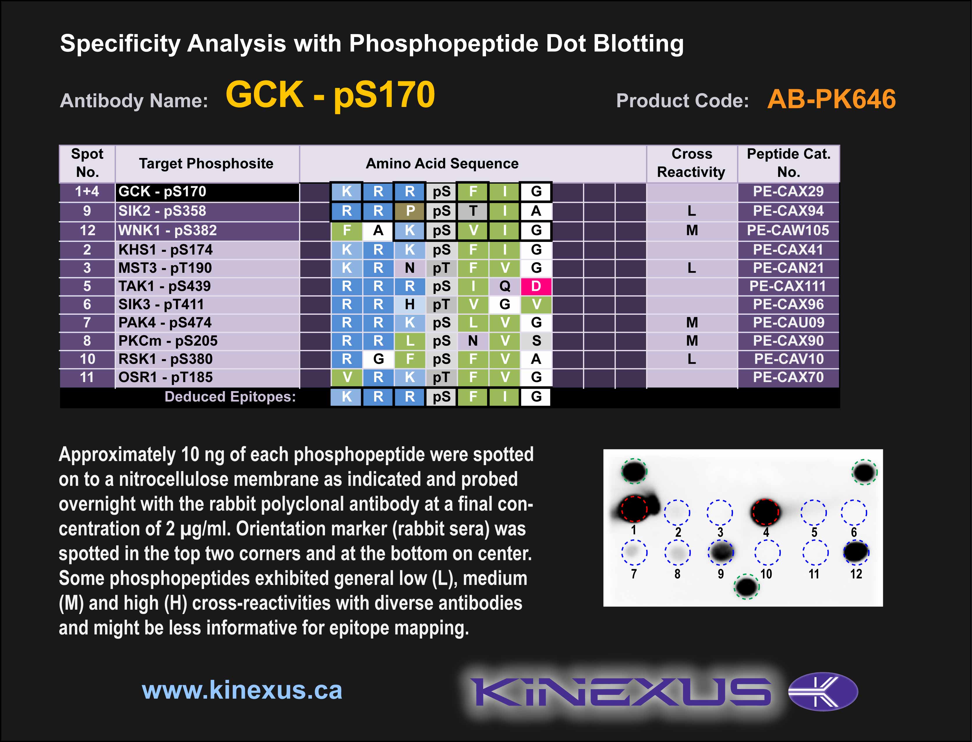
Figure 1. Epitope mapping of GCK-pS170 antibody with similar phosphopeptides on dot blots.
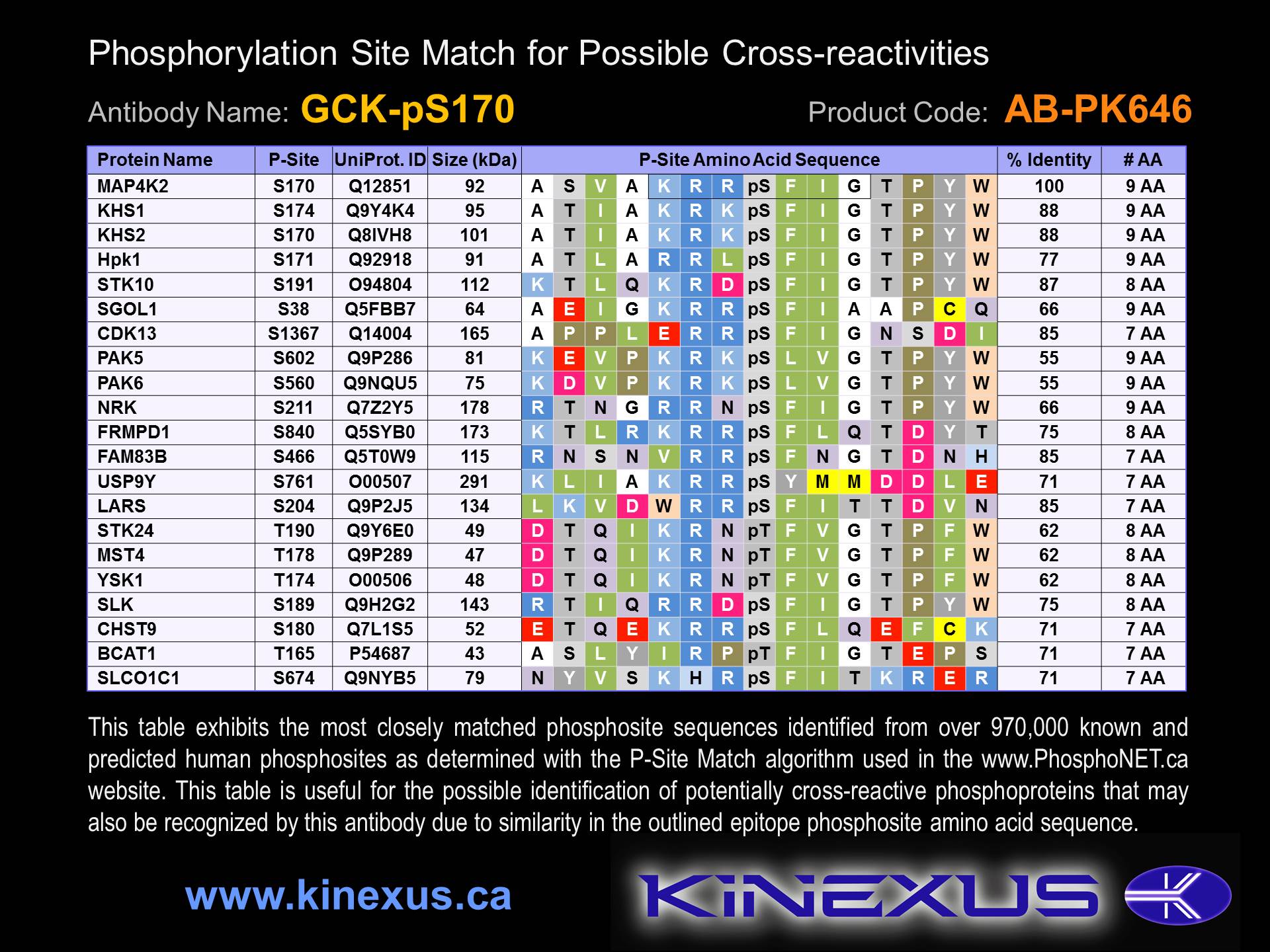
Figure 2. Identification of phosphosites related to GCK-pS170.

© Kinexus Bioinformatics Corporation 2017