
Product Name: HIPK1-pY352
Product Number: AB-PK654
| Size: | 25 µg | Price: | 89.00 | |
| $US |
Target Full Name: Homeodomain-interacting protein-serine kinase 1
Target Alias: HIK1; Homeodomain interacting protein kinase 1; KIAA0630; MGC26642; MGC33446; MGC33548; Myak; Nbak2
Product Type Specific: Protein kinase phosphosite-specific antibody
Antibody Code: PK654
Antibody Target Type: Phosphosite-specific
Antibody Phosphosite: Y352
Protein UniProt: Q86Z02
Protein SigNET: Q86Z02
Antibody Type: Polyclonal
Antibody Host Species: Rabbit
Antibody Immunogen Source: Human HIPK1 sequence peptide Cat. No.: PE-04AKK95
Antibody Immunogen Sequence: CST(pY)LQS(bA)C
Antibody Immunogen Description: Corresponds to amino acid residues C349 to S355; In protein kinase catalytic domain activation T-loop between subdomains VII and VIII. This is the major in vivo phosphorylation site in HIPK1.
Antibody Immunogen Description: Corresponds to amino acid residues C349 to S355; In protein kinase catalytic domain activation T-loop between subdomains VII and VIII. This is the major in vivo phosphorylation site in HIPK1.
Production Method: The immunizing peptide was produced by solid phase synthesis on a multipep peptide synthesizer and purified by reverse-phase hplc chromatography. Purity was assessed by analytical hplc and the amino acid sequence confirmed by mass spectrometry analysis. This peptide was coupled to KLH prior to immunization into rabbits. New Zealand White rabbits were subcutaneously injected with KLH-coupled immunizing peptide every 4 weeks for 4 months. The sera from these animals was applied onto an agarose column to which the immunogen peptide was thio-linked. Antibody was eluted from the column with 0.1 M glycine, pH 2.5. Subsequently, the antibody solution was neutralized to pH 7.0 with saturated Tris.This antibody was also subject to negative purification over phosphotyrosine-agarose.
Antibody Modification: Unconjugated. Contact KInexus if you are interest in having the antibody biotinylated or coupled with fluorescent dyes.
Antibody Modification: Unconjugated. Contact KInexus if you are interest in having the antibody biotinylated or coupled with fluorescent dyes.
Antibody Concentration: 1 mg/ml
Storage Buffer: Phosphate buffered saline pH 7.4, 0.05% Thimerasol
Storage Conditions: For long term storage, keep frozen at -40°C or lower. Stock solution can be kept at +4°C for more than 3 months. Avoid repeated freeze-thaw cycles.
Product Use: Western blotting | Antibody microarray
Antibody Dilution Recommended: 2 µg/ml for immunoblotting
Antibody Potency: Medium immunoreactivity of a target-sized protein by Western blotting in maturing, germinal vesicle positive, sea star oocytes. Very strong immunoreactivity with immunogen peptide on dot blots.
Antibody Species Reactivity: Human
Antibody Positive Control: The observed molecular mass of the processed target protein on SDS-PAGE gels is reported to be around 125-135 kDa.
Antibody Specificity: High
Antibody Cross Reactivity: No significant cross-reactive proteins detected in T98G cells. Strong 40 and 46 kDa cross-reactive proteins were detected in MCF7 cells and strong 22 kDa cross-reactive proteins in HeLa cells.
Related Product 1: HIPK1-pY352 blocking peptide
Related Product 2: HIPK2-BCT pan-specific antibody (Cat. No.: AB-NK272-1)
Related Product 3: DYRKtide KinSub - DYR peptide substrate
Scientific Background: HIPK1 (HIK1) is a protein-serine/threonine kinase of the CMGC group and DYRK family. It functions in transcription regulation as well as tumour necrosis factor (TNF)-mediated apoptosis. HIPK1 is also known to act as a co-repressor for homeodomain transcription factors. In the absence of TNF, HIPK1 prevents MAP3K5-JNK activation. When present, TNF induces nuclear translocation of HIPK1, thus removing it from the cytoplasm, resulting in the activation of MAP3K5-JNK signalling by de-repression. In addition, through the direct phosphorylation of p53 at the S15 residue, HIPK1 activates p53 leading to increased p21 protein levels, which subsequently inhibits cell proliferation. HIPK1 may be a tumour suppressor protein (TSP). Over-expression of HIPK1 has been observed in colorectal cancer specimens, with highest levels at the beginning of tumourigenesis followed by a progressive reduction over time as the cancer develops, indicating a role for the HIPK1 protein in the early events of tumourigenesis. in addition, these specimens also displayed native p53 and normal levels of p21, indicating that the HIPK1 protein functions as a tumour suppressor and that a loss-of-function in the protein is associated with tumourigenesis.
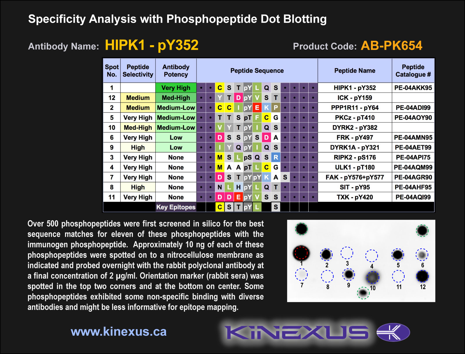
Figure 1. Epitope mapping of HIPK1-pY352 antibody with similar phosphopeptides on dot blots.
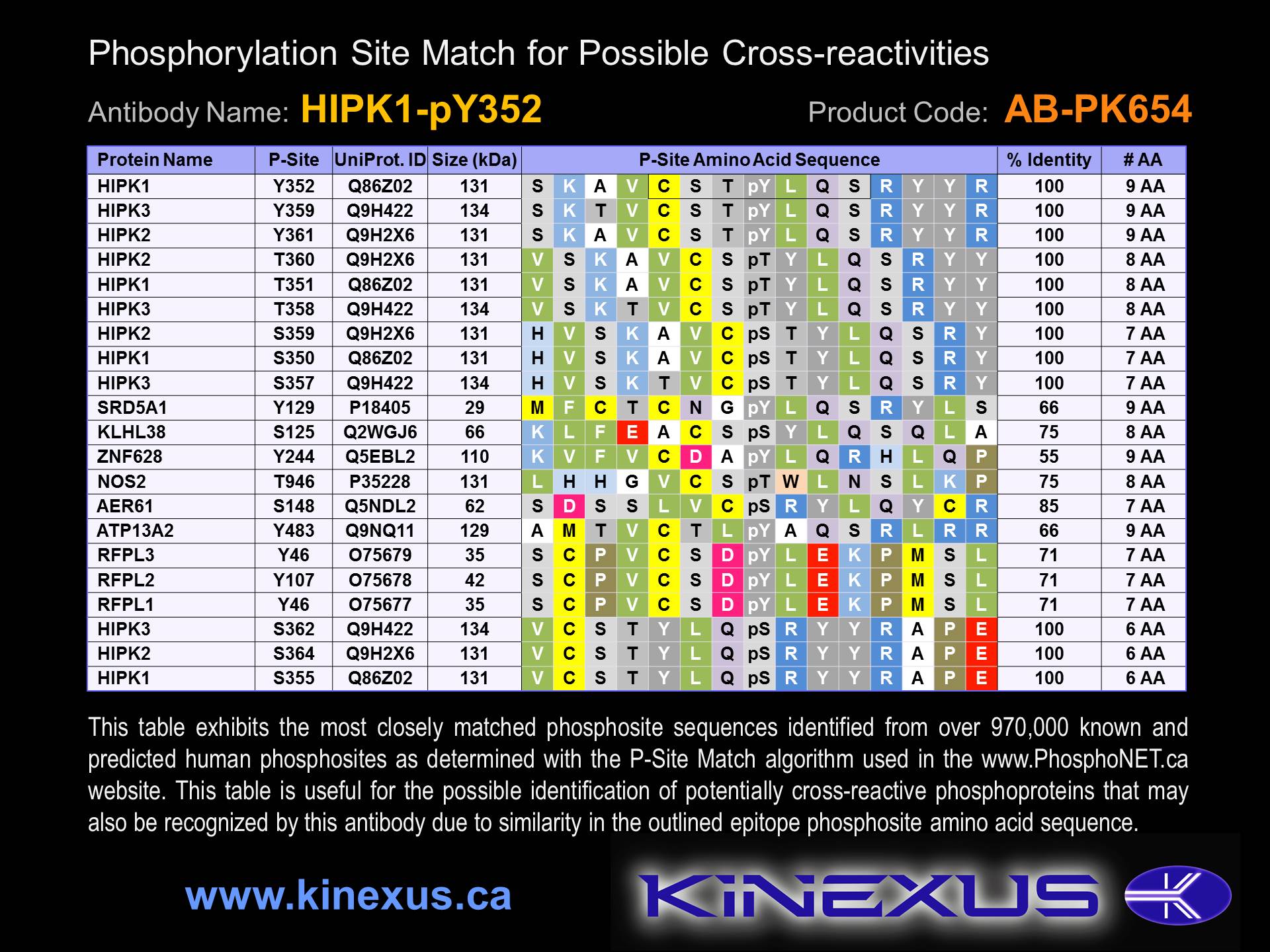
Figure 2. Identification of phosphosites related to HIPK1-pY352.

© Kinexus Bioinformatics Corporation 2017