
Product Name: ILK1-pS343
Product Number: AB-PK661
| Size: | 25 µg | Price: | 89.00 | |
| $US |
Target Full Name: Integrin-linked protein-serine kinase-1
Target Alias: 59 kDa serine/threonine protein kinase; ILK1; ILK-1; Integrin-linked kinase; Kinase ILK; P59ILK; ILK2; DKFZp686F1765; P59; CCDS7768.1; ENSG00000166333
Product Type Specific: Protein kinase phosphosite-specific antibody
Antibody Code: PK661
Antibody Target Type: Phosphosite-specific
Antibody Phosphosite: S343
Protein UniProt: Q13418
Protein SigNET: Q13418
Antibody Type: Polyclonal
Antibody Host Species: Rabbit
Antibody Immunogen Source: Human ILK1 sequence peptide Cat. No.: PE-04AMU99
Antibody Immunogen Sequence: DVKF(pS)FQC
Antibody Immunogen Description: Corresponds to amino acid residues D339 to C346; In the protein kinase catalytic domain activation T loop region between subdomains VII and VIII.
Antibody Immunogen Sequence: DVKF(pS)FQC
Antibody Immunogen Description: Corresponds to amino acid residues D339 to C346; In the protein kinase catalytic domain activation T loop region between subdomains VII and VIII.
Production Method: The immunizing peptide was produced by solid phase synthesis on a multipep peptide synthesizer and purified by reverse-phase hplc chromatography. Purity was assessed by analytical hplc and the amino acid sequence confirmed by mass spectrometry analysis. This peptide was coupled to KLH prior to immunization into rabbits. New Zealand White rabbits were subcutaneously injected with KLH-coupled immunizing peptide every 4 weeks for 4 months. The sera from these animals was applied onto an agarose column to which the immunogen peptide was thio-linked. Antibody was eluted from the column with 0.1 M glycine, pH 2.5. Subsequently, the antibody solution was neutralized to pH 7.0 with saturated Tris.This antibody was also subject to negative purification over phosphotyrosine-agarose.
Antibody Modification: Unconjugated. Contact KInexus if you are interest in having the antibody biotinylated or coupled with fluorescent dyes.
Antibody Modification: Unconjugated. Contact KInexus if you are interest in having the antibody biotinylated or coupled with fluorescent dyes.
Antibody Concentration: 0.15 mg/ml
Storage Buffer: Phosphate buffered saline pH 7.4, 0.05% Thimerasol
Storage Conditions: For long term storage, keep frozen at -40°C or lower. Stock solution can be kept at +4°C for more than 3 months. Avoid repeated freeze-thaw cycles.
Product Use: Western blotting | Antibody microarray
Antibody Dilution Recommended: 2 µg/ml for immunoblotting
Antibody Potency: Medium immunoreactivity with immunogen peptide on dot blots.
Antibody Species Reactivity: Human
Antibody Positive Control: The observed molecular mass of the processed target protein on SDS-PAGE gels is reported to be around 50-55 kDa.
Antibody Specificity: Very high
Antibody Cross Reactivity: No significant cross-reactive proteins detected in phenylarsine oxide (PAO)+vanadate-treated HeLa cells, EGF-treated A431 cells and insulin-treated MCF7 cells, when these cells were homogenized in SDS-PAGE sample buffer.
Related Product 1: ILK1-pS343 blocking peptide
Related Product 2: ILK1-pY351 phosphosite-specific antibody (Cat. No.: AB-PK662)
Scientific Background: ILK (ILK1) is a protein-serine/threonine kinase of the TKL group and MLK family. It has a critical role in the regulation of integrin-mediated signal transduction. It is stimulated rapidly but transiently by both cell fibronectin interactions, as well as by insulin, in a PI3-K-dependent manner, likely via the binding of PtdIns(3,4,5)P3 with a PH-like domain of ILK. It is a component of the complex ILK-PINCH, which is thought to be a primary point for the convergence of integrin- and growth-factor signalling as well as a regulator of cell architecture, integrin-mediated adhesion, and anchorage-dependent cell growth in epithelial cells. Downstream targets of ILK include the beta-1 and beta-3 integrin subunits as well as Akt1 and GSK3b. The ILK mutation, H99D can modulate interactions with LIMS1. An E359K mutation can inactivate ILK. ILK appears to be a oncoprotein (OP), but is relatively low rate of mutation in human cancers supports its assignment as a tumour requiring protein (TRP). It has been implicated as an oncoprotein due to its ability to activate Akt isoforms, although it has weak phosphotransferase activity that is stimulated in vitro with manganese.
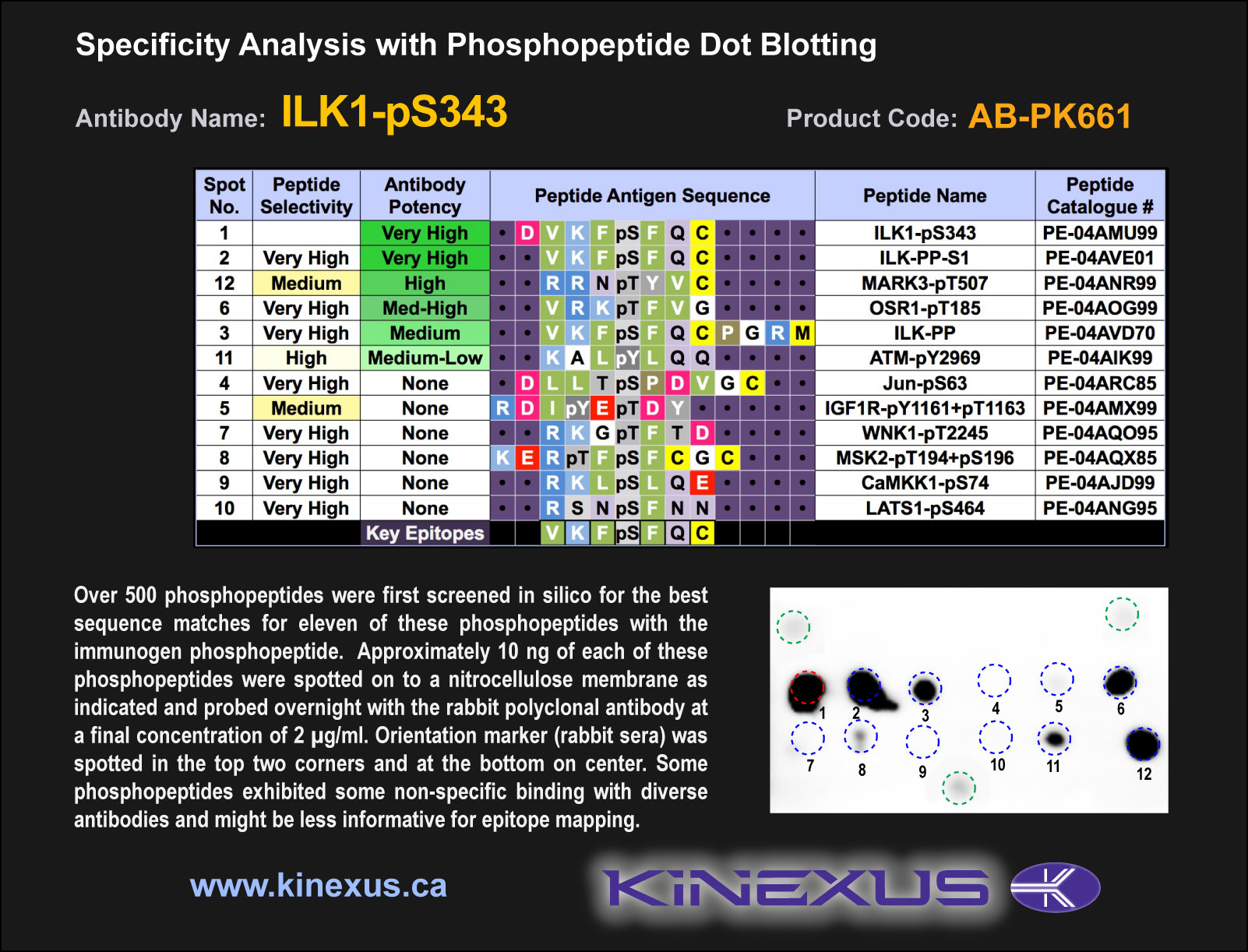
Figure 1. Epitope mapping of ILK1-pS343 antibody with similar phosphopeptides on dot blots.
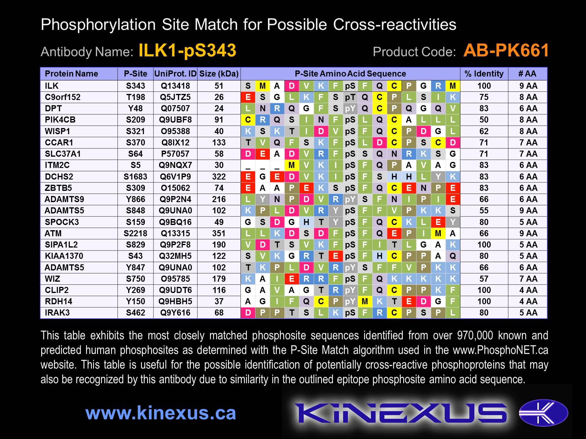
Figure 2. Identification of phosphosites related to ILK1-pS343.

© Kinexus Bioinformatics Corporation 2017