
Product Name: IRAK1-pT387
Product Number: AB-PK664
| Size: | 25 µg | Price: | 89.00 | |
| $US |
Target Full Name: Interleukin 1 receptor-associated kinase 1 (Pelle-like protein kinase)
Target Alias: IL1RAK; IRAK; IRAK-1; Kinase IRAK1; MPLK; Pelle; Pelle-like protein kinase
Product Type Specific: Protein kinase phosphosite-specific antibody
Antibody Code: PK664
Antibody Target Type: Phosphosite-specific
Antibody Phosphosite: T387
Protein UniProt: P51617
Protein SigNET: P51617
Antibody Type: Polyclonal
Antibody Host Species: Rabbit
Antibody Immunogen Source: Human IRAK1 sequence peptide Cat. No.: PE-04AMY95
Antibody Immunogen Sequence: VRG(pT)LAY(bA)C
Antibody Immunogen Description: Corresponds to amino acid residues V384 to Y390; In protein kinase catalytic domain activation T-loop between subdomains VII and VIII.
Antibody Immunogen Sequence: VRG(pT)LAY(bA)C
Antibody Immunogen Description: Corresponds to amino acid residues V384 to Y390; In protein kinase catalytic domain activation T-loop between subdomains VII and VIII.
Production Method: The immunizing peptide was produced by solid phase synthesis on a multipep peptide synthesizer and purified by reverse-phase hplc chromatography. Purity was assessed by analytical hplc and the amino acid sequence confirmed by mass spectrometry analysis. This peptide was coupled to KLH prior to immunization into rabbits. New Zealand White rabbits were subcutaneously injected with KLH-coupled immunizing peptide every 4 weeks for 4 months. The sera from these animals was applied onto an agarose column to which the immunogen peptide was thio-linked. Antibody was eluted from the column with 0.1 M glycine, pH 2.5. Subsequently, the antibody solution was neutralized to pH 7.0 with saturated Tris.This antibody was also subject to negative purification over phosphotyrosine-agarose.
Antibody Modification: Unconjugated. Contact KInexus if you are interest in having the antibody biotinylated or coupled with fluorescent dyes.
Antibody Modification: Unconjugated. Contact KInexus if you are interest in having the antibody biotinylated or coupled with fluorescent dyes.
Antibody Concentration: 0.5 mg/ml
Storage Buffer: Phosphate buffered saline pH 7.4, 0.05% Thimerasol
Storage Conditions: For long term storage, keep frozen at -40°C or lower. Stock solution can be kept at +4°C for more than 3 months. Avoid repeated freeze-thaw cycles.
Product Use: Western blotting | Antibody microarray
Antibody Dilution Recommended: 2 µg/ml for immunoblotting
Antibody Potency: Medium immunoreactivity with immunogen peptide on dot blots.
Antibody Species Reactivity: Human
Antibody Positive Control: The observed molecular mass of the processed target protein on SDS-PAGE gels is reported to be around 72-80 kDa.
Antibody Specificity: High-very high
Antibody Cross Reactivity: No significant cross-reactive proteins detected in phenylarsine oxide (PAO)+vanadate-treated HeLa cells, EGF-treated A431 cells and insulin-treated MCF7 cells, when these cells were homogenized in SDS-PAGE sample buffer, except for a 26K cross-reactive protein in EGF-treated HeLa cells.
Related Product 1: IRAK1-pT387 blocking peptide
Related Product 2: IRAKSelectide - IRAK1 protein kinase substrate peptide
Scientific Background: IRAK1 is a protein-serine/threonine kinase of the TKL group and IRAK family. It plays a critical role in initiating innate immune response against foreign pathogens. It is involved in Toll-like receptor (TLR) and IL-1R signalling pathways, and Is rapidly recruited by MYD88 to the receptor-signalling complex upon TLR activation. Its association with MYD88 leads to IRAK1 phosphorylation by IRAK4 and subsequent autophosphorylation and kinase activation. It phosphorylates E3 ubiquitin ligases Pellino proteins (PELI1, PELI2 and PELI3) to promote pellino-mediated polyubiquitination of IRAK1. Then, the ubiquitin-binding domain of IKBKG/NEMO binds to polyubiquitinated IRAK1 bringing together the IRAK1-MAP3K7/TAK1-TRAF6 complex and the NEMO-IKKA-IKKB complex. IRAK1 phosphorylates TIRAP to promote its ubiquitination and subsequent degradation. It phosphorylates the interferon regulatory factor 7 (IRF7) to induce its activation and translocation to the nucleus, to stimulate transcriptional activation of type I IFN genes, which drive the cell in an antiviral state. When sumoylated, IRAK1 translocates to the nucleus and phosphorylates STAT3. This kinase is moderate to highly expressed in most tested human tissues.
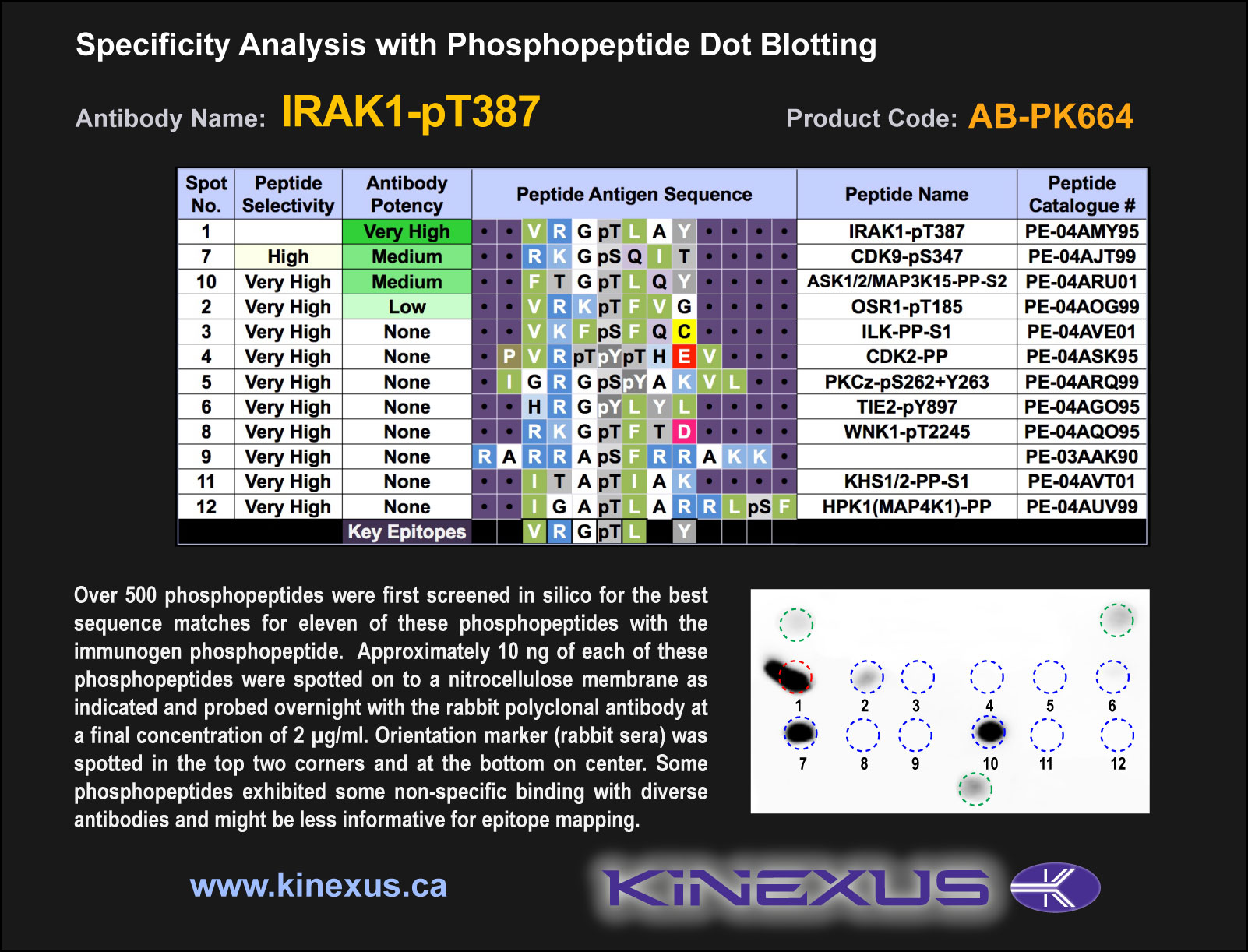
Figure 1. Epitope mapping of IRAK1-pT387 antibody with similar phosphopeptides on dot blots.
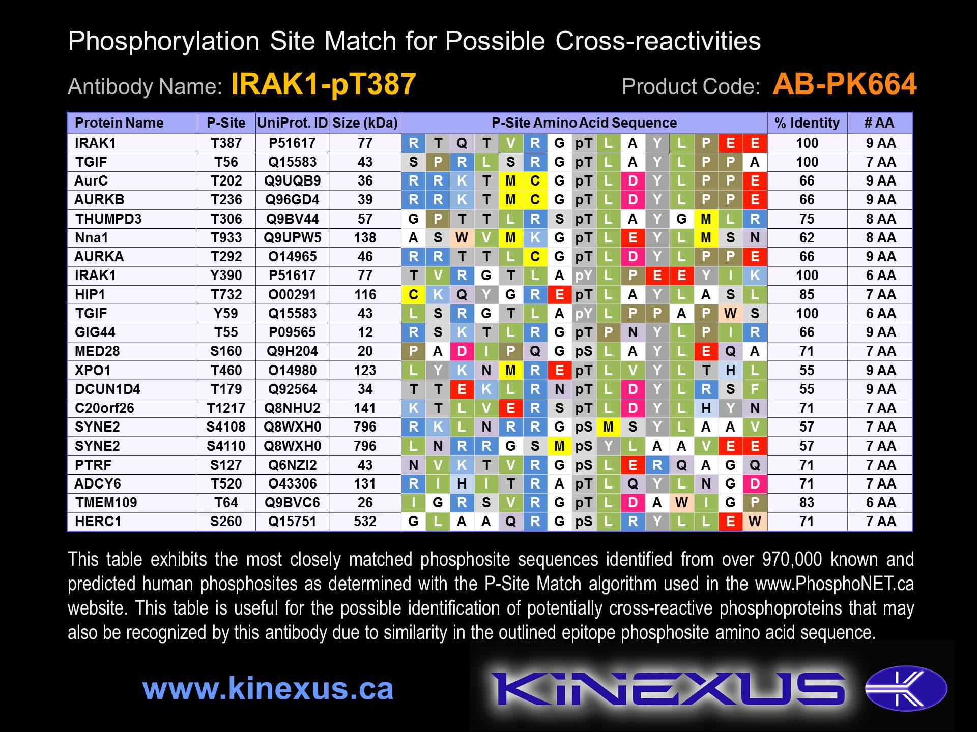
Figure 2. Identification of phosphosites related to IRAK1-pT387.

© Kinexus Bioinformatics Corporation 2017