
Product Name: MST3-pT184
Product Number: AB-PK727
| Size: | 25 µg | Price: | 89.00 | |
| $US |
Target Full Name: Mammalian STE20-like protein-serine kinase 3; Serine-threonine-protein kinase 24
Target Alias: MST3 isoform B; MST3b; RP11-111L24.5; Serine,threonine protein kinase 24; STE20-like kinase MST3; STK24; STK3
Product Type Specific: Protein kinase phosphosite-specific antibody
Antibody Code: PK727
Antibody Target Type: Phosphosite-specific
Antibody Phosphosite: T184
Protein UniProt: Q9Y6E0
Protein SigNET: Q9Y6E0
Antibody Type: Polyclonal
Antibody Host Species: Rabbit
Antibody Immunogen Source: Human MST3 (STK24) sequence peptide Cat. No.: PE-04ABK90
Antibody Immunogen Sequence: LTD(pT)QIK(bA)C
Antibody Immunogen Description: Corresponds to amino acid residues L181 to K187; In protein kinase catalytic domain activation T-loop between subdomains VII and VIII.
Antibody Immunogen Sequence: LTD(pT)QIK(bA)C
Antibody Immunogen Description: Corresponds to amino acid residues L181 to K187; In protein kinase catalytic domain activation T-loop between subdomains VII and VIII.
Production Method: The immunizing peptide was produced by solid phase synthesis on a multipep peptide synthesizer and purified by reverse-phase hplc chromatography. Purity was assessed by analytical hplc and the amino acid sequence confirmed by mass spectrometry analysis. This peptide was coupled to KLH prior to immunization into rabbits. New Zealand White rabbits were subcutaneously injected with KLH-coupled immunizing peptide every 4 weeks for 4 months. The sera from these animals was applied onto an agarose column to which the immunogen peptide was thio-linked. Antibody was eluted from the column with 0.1 M glycine, pH 2.5. Subsequently, the antibody solution was neutralized to pH 7.0 with saturated Tris.This antibody was also subject to negative purification over phosphotyrosine-agarose.
Antibody Modification: Unconjugated. Contact KInexus if you are interest in having the antibody biotinylated or coupled with fluorescent dyes.
Antibody Modification: Unconjugated. Contact KInexus if you are interest in having the antibody biotinylated or coupled with fluorescent dyes.
Antibody Concentration: 1 mg/ml
Storage Buffer: Phosphate buffered saline pH 7.4, 0.05% Thimerasol
Storage Conditions: For long term storage, keep frozen at -40°C or lower. Stock solution can be kept at +4°C for more than 3 months. Avoid repeated freeze-thaw cycles.
Product Use: Western blotting | Antibody microarray
Antibody Dilution Recommended: 2 µg/ml for immunoblotting
Antibody Potency: Very strong immunoreactivity with immunogen peptide on dot blots.
Antibody Species Reactivity: Human
Antibody Positive Control: The observed molecular mass of the processed target protein on SDS-PAGE gels is reported to be around 50-55 kDa.
Antibody Specificity: Very high
Antibody Cross Reactivity: No significant cross-reactive proteins detected in T98G cells.
Related Product 1: MST3-pT184 blocking peptide
Related Product 2: MST3-pT190 phosphosite-specific antibody (Cat. No.: AB-PK728)
Related Product 3: IRS1 (979-989) KinSub - Insulin receptor substrate 1 (K979-G989, mouse) peptide; Insulin receptor substrate
Related Product 4: MstSubtide - MST1 (STK4) protein kinase substrate peptide
Scientific Background: MST3 (STK24) is a protein-serine/threonine kinase that is a member of the STE group of protein kinases in the STE20 family, and YSK subfamily. This kinase is highly expressed and widely distributed in most tested human tissues. MST3 promotes apoptosis in response to stress stimuli and caspase activation. It indirectly activates JNK1-JNK2 (MAPK8 and MAPK9), p38 (MAPK11, MAPK12, MAPK13 and MAPK14 ) to mediates oxidative-stress-induced apoptosis. MST3 contains a conserved kinase domain at its N-terminus and a regulatory domain at its C-terminus. Caspase-mediated cleavage of the regulatory domain of MST3 activates its intrinsic kinase activity and leads to its nuclear translocation. Expression of C-terminal truncated MST3 in cells results in DNA fragmentation and induction of apoptosis. MST3 can inhibit cell migration in a fashion dependent on autophosphorylation and can regulate paxillin phosphorylation through protein-tyrosine phosphatase PTP-PEST. It has been implicated a key regulator of axon regeneration in the optic nerve and radial nerve.
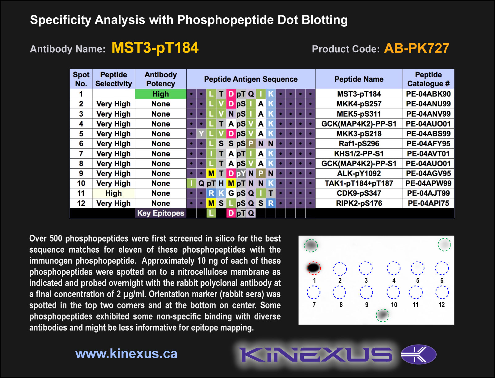
Figure 1. Epitope mapping of MST3-pT184 antibody with similar phosphopeptides on dot blots.
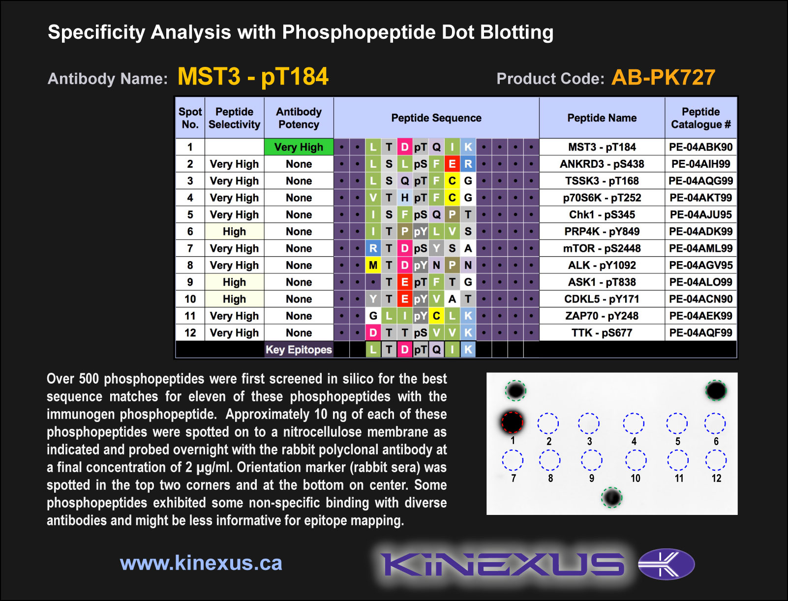
Figure 2. Epitope mapping of MST3-pT184 antibody with similar phosphopeptides on dot blots.
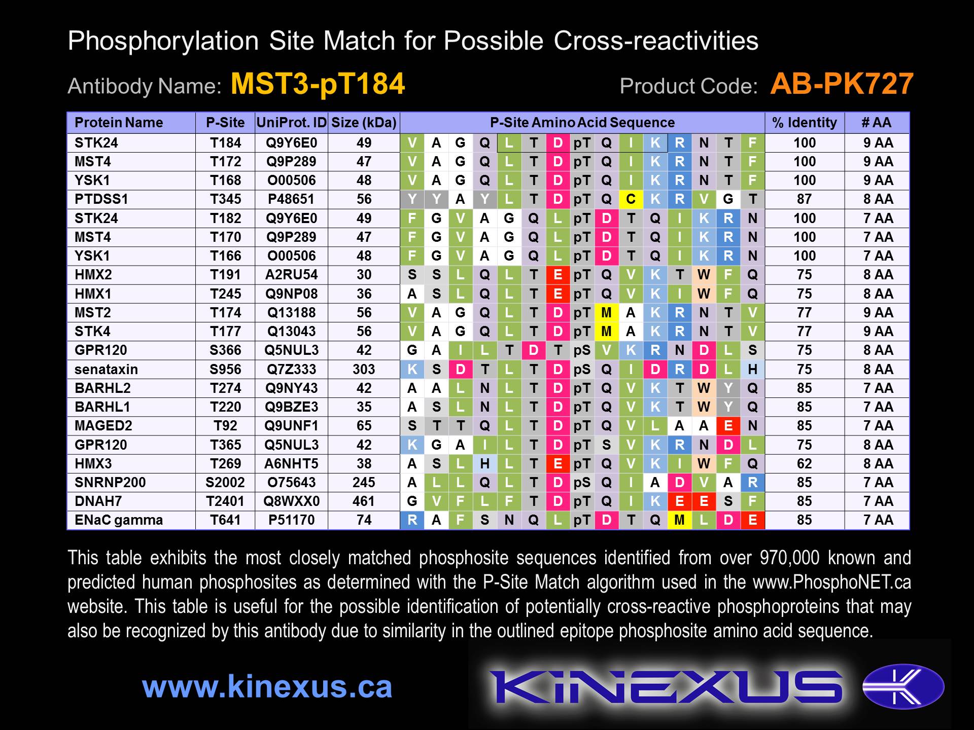
Figure 3. Identification of phosphosites related to MST3-pT184.

© Kinexus Bioinformatics Corporation 2017