
Product Name: PAK2-pS141
Product Number: AB-PK750
| Size: | 25 µg | Price: | 89.00 | |
| $US |
Target Full Name: p21-activated kinase 2 gamma; Protein-serine/threonine kinase PAK 2
Target Alias: C-t-PAK2; Gamma-PAK; Kinase PAK2; P21 protein (Cdc42/Rac)-activated kinase 2; P21-activated kinase 2; p21-activated protein kinase I; P58; PAK 2; PAK-2; S6/H4 kinase; PAK65; CCDS3321.1; Q6ISC3; ENSG00000180370
Product Type Specific: Protein kinase phosphosite-specific antibody
Antibody Code: PK750
Antibody Target Type: Phosphosite-specific
Antibody Phosphosite: S141
Protein UniProt: Q13177
Protein SigNET: Q13177
Antibody Type: Polyclonal
Antibody Host Species: Rabbit
Antibody Immunogen Source: Human PAK2 (PAKg) sequence peptide Cat. No.: PE-04AOJ99
Antibody Immunogen Sequence: KYL(pS)FTP(bA)C
Antibody Immunogen Description: Corresponds to amino acid residues K138 to P144; In the region between the PBD (p21-binding domain) and the kinase catalytic domain in the N-terminal third of the protein.
Production Method: The immunizing peptide was produced by solid phase synthesis on a multipep peptide synthesizer and purified by reverse-phase hplc chromatography. Purity was assessed by analytical hplc and the amino acid sequence confirmed by mass spectrometry analysis. This peptide was coupled to KLH prior to immunization into rabbits. New Zealand White rabbits were subcutaneously injected with KLH-coupled immunizing peptide every 4 weeks for 4 months. The sera from these animals was applied onto an agarose column to which the immunogen peptide was thio-linked. Antibody was eluted from the column with 0.1 M glycine, pH 2.5. Subsequently, the antibody solution was neutralized to pH 7.0 with saturated Tris.This antibody was also subject to negative purification over phosphotyrosine-agarose.
Antibody Modification: Unconjugated. Contact KInexus if you are interest in having the antibody biotinylated or coupled with fluorescent dyes.
Antibody Modification: Unconjugated. Contact KInexus if you are interest in having the antibody biotinylated or coupled with fluorescent dyes.
Antibody Concentration: 1 mg/ml
Storage Buffer: Phosphate buffered saline pH 7.4, 0.05% Thimerasol
Storage Conditions: For long term storage, keep frozen at -40°C or lower. Stock solution can be kept at +4°C for more than 3 months. Avoid repeated freeze-thaw cycles.
Product Use: Western blotting | Antibody microarray
Antibody Dilution Recommended: 2 µg/ml for immunoblotting
Antibody Potency: Very strong immunoreactivity of a target-sized protein by Western blotting in HepG2 and T98G cells, and human lung. Very strong immunoreactivity with immunogen peptide on dot blots.
Antibody Species Reactivity: Human
Antibody Positive Control: The observed molecular mass of the processed target protein on SDS-PAGE gels is reported to be around 60-67 kDa.
Antibody Specificity: High-very high
Antibody Cross Reactivity: Strong cross-reactive ~52 and 48 kDa proteins in human lung. No significant cross-reactivities detected in HepG2 and T98G cells.
Related Product 1: PAK2-pS141 blocking peptide
Related Product 2: PAK2-NT (PAK2-1) pan-specific antibody (Cat. No.: AB-NK200)
Related Product 3: PAK2-pY130 phosphosite-specific antibody (Cat. No.: AB-PK751)
Related Product 4: PAktide KinSub - PAK peptide substrate
Scientific Background: PAK2 (PAK65) is a protein-serine/threonine kinase of the STE group and STE20 family. It functions in the regulation of several cellular processes including cytoskeletal reorganization, cell motility, cell cycle progression, apoptosis, and cellular proliferation. Under conditions that promotes cell growth and survival, PAK2 is activated by binding small G proteins. Binding of GTP-bound Cdc42 or Rac1 to the autoregulatory region releases monomers from the autoinhibited dimer, enables phosphorylation of T402 and allows the kinase domain to adopt an active structure. Phosphorylation at Y130, S141 and T402 increases phosphotransferase activity. It phosphorylates MAPK4 and MAPK6 ,c-Jun, histone H4, BAD, ribosomal protein S6, and MBP. Additionally, it associates with ARHGEF7 and GIT1 to perform kinase-independent functions such as spindle orientation control during mitosis. Apoptotic stimuli (e.g. DNA damage) induce the cleavage of PAK2 by caspases, generating the constitutively active p34 fragment that subsequently translocates to the nucleus and stimulates apoptosis via the JNK signalling pathway and phosphorylates MKNK1 to reduces cellular translation. PAK2 may be an oncoprotein (OP). Significantly elevated S20 phosphorylated PAK2 was observed in ovarian cancer cell lines. In addition, knockdown of PAK2 expression in ovarian cancer cell lines reduces cell migration and invasion without affecting cell proliferation or apoptosis. Elevated PAK2 and pPAK2 expression have also been demonstrated in human gastric cancer cell lines. Furthermore, elevated PAK2 and pPAK2 expression have been correlated with several unfavourable variables in patients with gastric cancer including higher tumour depth, greater extent of metastasis, advanced tumour stage and significantly lower survival rates.
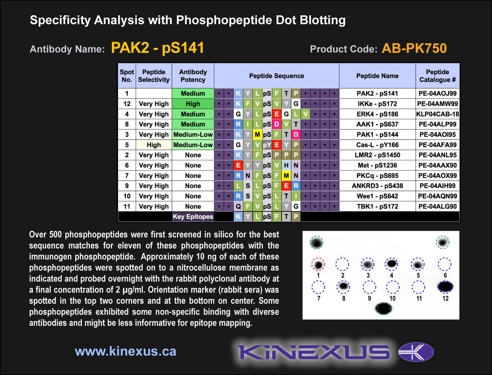
Figure 1. Epitope mapping of PAK2-pS141 antibody with similar phosphopeptides on dot blots.
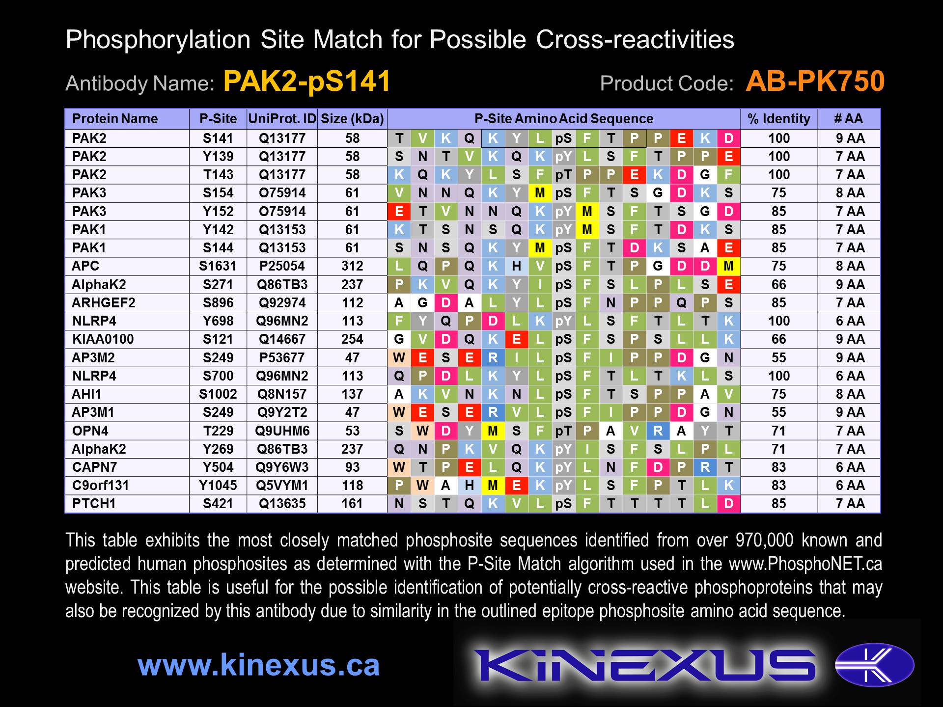
Figure 2. Identification of phosphosites related to PAK2-pS141.

© Kinexus Bioinformatics Corporation 2017