
Product Name: PAK5-pS602
Product Number: AB-PK753
| Size: | 25 µg | Price: | 89.00 | |
| $US |
Target Full Name: p21-activated kinase 5; Protein-serine/threonine kinase PAK 7
Target Alias: KIAA1264; P21 protein (Cdc42/Rac)-activated kinase 7; P21-activated kinase 7; PAK 7; PAK-5; PAK7; PAK-7; MGC26232; RP5-1119D9_3; ENSG00000101349; KIAA1264
Product Type Specific: Protein kinase phosphosite-specific antibody
Antibody Code: PK753
Antibody Target Type: Phosphosite-specific
Antibody Phosphosite: S602
Protein UniProt: Q9P286
Protein SigNET: Q9P286
Antibody Type: Polyclonal
Antibody Host Species: Rabbit
Antibody Immunogen Source: Human PAK5 (PAK7) sequence peptide Cat. No.: PE-04AOL99
Antibody Immunogen Sequence: KRK(pS)LVG(bA)C
Antibody Immunogen Description: Corresponds to amino acid residues K599 to G605; In the protein kinase catalytic domain activation T loop region between subdomains VII and VIII.
Antibody Immunogen Sequence: KRK(pS)LVG(bA)C
Antibody Immunogen Description: Corresponds to amino acid residues K599 to G605; In the protein kinase catalytic domain activation T loop region between subdomains VII and VIII.
Production Method: The immunizing peptide was produced by solid phase synthesis on a multipep peptide synthesizer and purified by reverse-phase hplc chromatography. Purity was assessed by analytical hplc and the amino acid sequence confirmed by mass spectrometry analysis. This peptide was coupled to KLH prior to immunization into rabbits. New Zealand White rabbits were subcutaneously injected with KLH-coupled immunizing peptide every 4 weeks for 4 months. The sera from these animals was applied onto an agarose column to which the immunogen peptide was thio-linked. Antibody was eluted from the column with 0.1 M glycine, pH 2.5. Subsequently, the antibody solution was neutralized to pH 7.0 with saturated Tris.This antibody was also subject to negative purification over phosphotyrosine-agarose.
Antibody Modification: Unconjugated. Contact KInexus if you are interest in having the antibody biotinylated or coupled with fluorescent dyes.
Antibody Modification: Unconjugated. Contact KInexus if you are interest in having the antibody biotinylated or coupled with fluorescent dyes.
Antibody Concentration: 1 mg/ml
Storage Buffer: Phosphate buffered saline pH 7.4, 0.05% Thimerasol
Storage Conditions: For long term storage, keep frozen at -40°C or lower. Stock solution can be kept at +4°C for more than 3 months. Avoid repeated freeze-thaw cycles.
Product Use: Western blotting | Antibody microarray
Antibody Dilution Recommended: 2 µg/ml for immunoblotting
Antibody Potency: Very strong immunoreactivity with immunogen peptide on dot blots.
Antibody Species Reactivity: Human
Antibody Positive Control: The observed molecular mass of the processed target protein on SDS-PAGE gels is reported to be around 75-85 kDa.
Related Product 1: PAK5-pS602 blocking peptide
Related Product 2: PAK5-ANT pan-specific antibody (Cat. No.: AB-NK190-2)
Related Product 3: PAK5-APBD pan-specific antibody (Cat. No.: AB-NK190-3)
Related Product 4: GSK3b (4-12) T7A, T8A; KinSub - Glycogen synthase kinase-3-beta-derived N-terminus (R4-E12, T7A, T8A, human) peptide; Akt1 protein kinase substrate peptide
Scientific Background: PAK5 (also known as PAK7) is a protein-serine/threonine that is a member of the STE group of protein kinases in the STE20 family, and PAKB subfamily. This kinase appears to have a restricted human tissue expression with the highest levels detected in brain, spinal cord and the adrenal gland. PAK5 has been implicated in the regulation of cell morphology, motility and transformation. PAK5 features a CDC42/Rac1 interactive binding (CRIB) motif at the N-terminus and a Ste20-like kinase domain at the C-terminus. Binding of GTP-bound Cdc42 or Rac1 to the autoregulatory region at the N-terminus of PAK5 releases monomers from the autoinhibited dimer, enables activation of its phosphotransferase activity. It phosphorylates BAD and prevents apoptosis. It is also involved in microtuble stablization, neurite outgrowth and development. It can destablize F-acting networks and eliminate stress fibres and focal adhesions. PAK5 expression provides a survival signal and could contribute to oncogenesis in this way. PAK5 is overexpressed in epithelial ovarian cancer (EOC), which is faced with an obstacle of paclitaxel resistance (PubMed: 23877225). Its expression was increased with EOC progression through the adenoma to carcinoma sequence, with the highest expression level of PAK5 in invasive and metastatic EOCs. Since PAK5 is correlated to human EOC, and its increased expression promotes EOC progression and induces EOC cell paclitaxel chemoresistance, PAK5 may be a useful target for therapeutic drug development. PAK5 has also been linked with the development of colorectal adenocarcinomas, lung adenocarcinomas, and melanomas (metastatic).
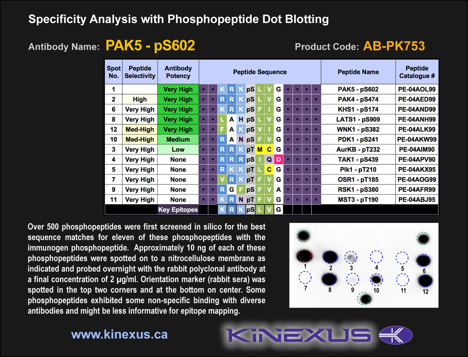
Figure 1. Epitope mapping of PAK5-pS602 antibody with similar phosphopeptides on dot blots.
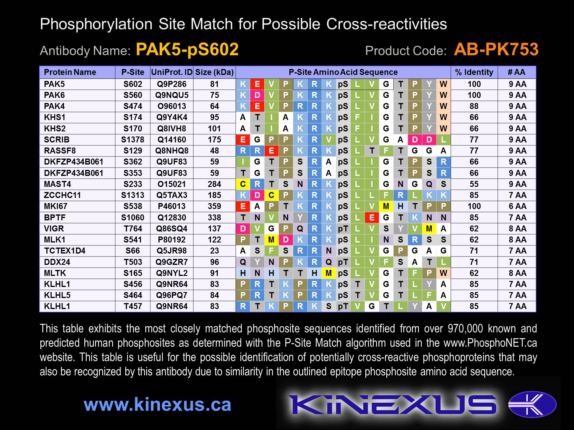
Figure 2. Identification of phosphosites related to PAK5-pS602.

PAK5-pS602

© Kinexus Bioinformatics Corporation 2017