
Product Name: PBK-pY74
Product Number: AB-PK754
| Size: | 25 µg | Price: | 89.00 | |
| $US |
Target Full Name: Lymphokine-activated killer T-cell-originated protein kinase
Target Alias: CT84; FLJ14385; NORI3; SPK; T-LAK cell-originated protein kinase; TOPK; TPOK
Product Type Specific: Protein kinase phosphosite-specific antibody
Antibody Code: PK754
Antibody Target Type: Phosphosite-specific
Antibody Phosphosite: Y74
Protein UniProt: Q96KB5
Protein SigNET: Q96KB5
Antibody Type: Polyclonal
Antibody Host Species: Rabbit
Antibody Immunogen Source: Human PBK sequence peptide Cat. No.: PE-04AOM95
Antibody Immunogen Sequence: NDH(pY)RSV(bA)C
Antibody Immunogen Description: Corresponds to amino acid residues N71 to V77; In the protein kinase catalytic domain between subdomains II and III. This is the major in vivo phosphorylation site in PBK.
Antibody Immunogen Description: Corresponds to amino acid residues N71 to V77; In the protein kinase catalytic domain between subdomains II and III. This is the major in vivo phosphorylation site in PBK.
Production Method: The immunizing peptide was produced by solid phase synthesis on a multipep peptide synthesizer and purified by reverse-phase hplc chromatography. Purity was assessed by analytical hplc and the amino acid sequence confirmed by mass spectrometry analysis. This peptide was coupled to KLH prior to immunization into rabbits. New Zealand White rabbits were subcutaneously injected with KLH-coupled immunizing peptide every 4 weeks for 4 months. The sera from these animals was applied onto an agarose column to which the immunogen peptide was thio-linked. Antibody was eluted from the column with 0.1 M glycine, pH 2.5. Subsequently, the antibody solution was neutralized to pH 7.0 with saturated Tris.This antibody was also subject to negative purification over phosphotyrosine-agarose.
Antibody Modification: Unconjugated. Contact KInexus if you are interest in having the antibody biotinylated or coupled with fluorescent dyes.
Antibody Modification: Unconjugated. Contact KInexus if you are interest in having the antibody biotinylated or coupled with fluorescent dyes.
Antibody Concentration: 0.5 / 1.0 mg/ml
Storage Buffer: Phosphate buffered saline pH 7.4, 0.05% Thimerasol
Storage Conditions: For long term storage, keep frozen at -40°C or lower. Stock solution can be kept at +4°C for more than 3 months. Avoid repeated freeze-thaw cycles.
Product Use: Western blotting | Antibody microarray
Antibody Dilution Recommended: 2 µg/ml for immunoblotting
Antibody Potency: Very strong immunoreactivity with immunogen peptide on dot blots.
Antibody Species Reactivity: Human
Antibody Positive Control: The observed molecular mass of the processed target protein on SDS-PAGE gels is reported to be around 36-44 kDa.
Antibody Specificity: Very high
Antibody Cross Reactivity: No significant cross-reactive proteins detected in phenylarsine oxide (PAO)+vanadate-treated HeLa cells, EGF-treated A431 cells and insulin-treated MCF7 cells, when these cells were homogenized in SDS-PAGE sample buffer, although strong background >75 kDa was observed.
Related Product 1: PBK-pY74 blocking peptide
Scientific Background: PBK (also known as TOPK) is a protein-serine/threonine kinase that is a member of the Other group of protein kinases in the TOPK family. This kinase shows high variability in human tissue distribution with the highest levels detected in lymph nodes, pituitary, spleen, testes and tonsils. Expression of PKB is particularly detected in male germ line progenitor cells, activated T-cells, and a variety of lymphomas and leukemias. T9 phosphorylation of PBK by CDK1 increases its phosphotransferase activity and induces interaction with CCNB1, CDK1, and tubulin-alpha1. PASK phosphorylates p38 MAPK (but not JNK or ERK1/2) and is activated in a cell-cycle-dependent manner in neuronal progenitor cells in vitro. PBK is active in mitosis and inhibition of the PBK/p38 pathway disrupts neural progentior proliferation and self renewal. In addition, PBK is hypothesized to function in the activation of lymphoid cells. When activated, PBK forms a complex with p53, which destablizes the p53 protein and leads to the attenuation of the G2/M checkpoint for DNA damage. PKB activation requires phosphorylation by both the M-phase CDK1/CyclinB kinase complex and another unknown kinase, possibly Raf1 or RafA. PKB may play an important role in linking extracellular signals to an intracellular state, possibly allowing extracellular influence on the cell-cycle-related processes of proliferation or differentiation. PBK may be an oncoprotein (OP). Gain-of-function mutations in PBK are linked to cancer, specifically plexiform neuroblastoma, as an overactive PBK protein impairs the G2/M checkpoint and prevents cell cycle arrest in the presence of DNA damage. Despite being absent from most normal somatic tissues, PBK has been shown to be significantly upregulated in several cancer types, indicating that gain-of-function mutations may be causing aberrant PBK expression leading to the augmentation of tumour growth in several forms of human cancer.
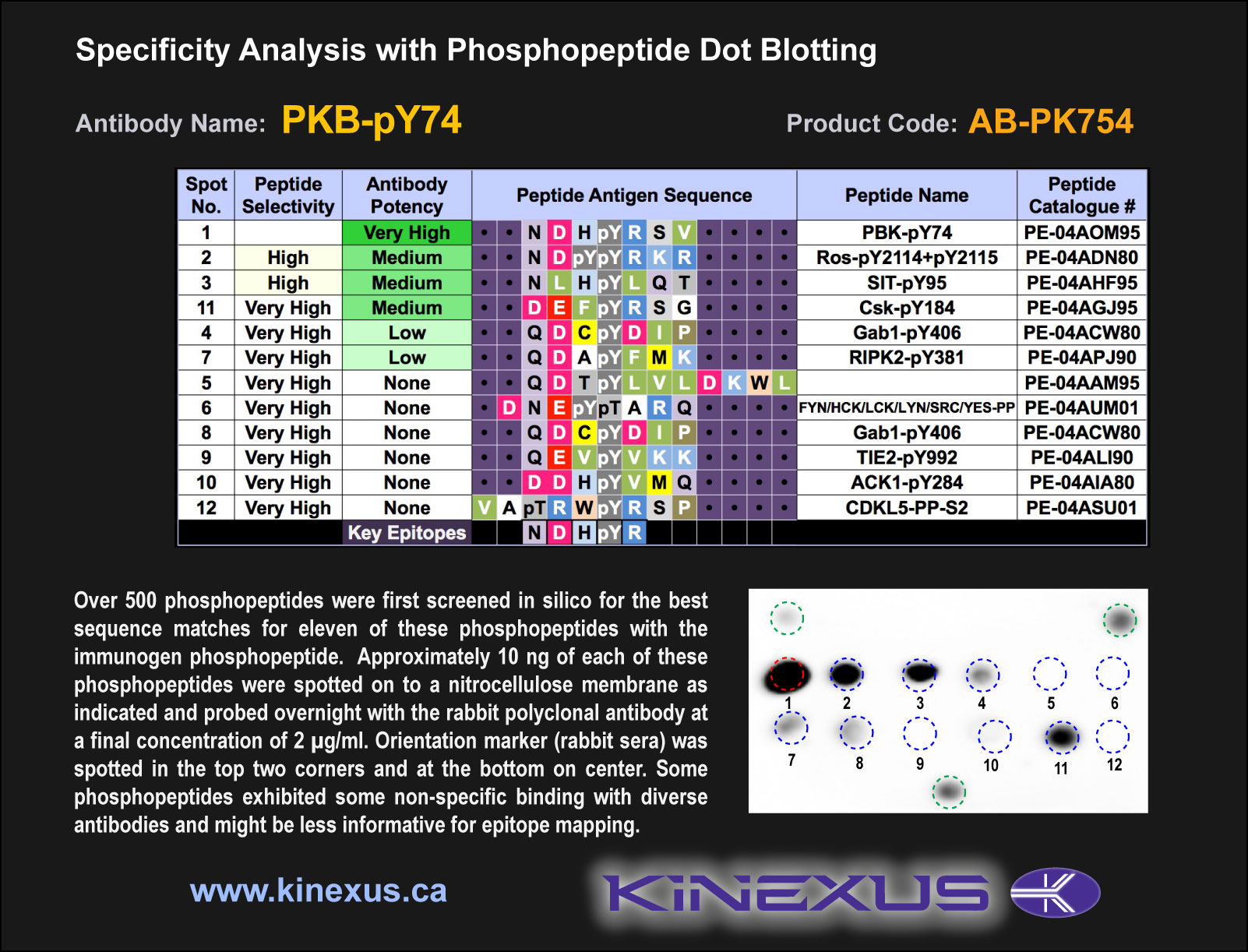
Figure 1. Epitope mapping of PBK-pY74 antibody with similar phosphopeptides on dot blots.
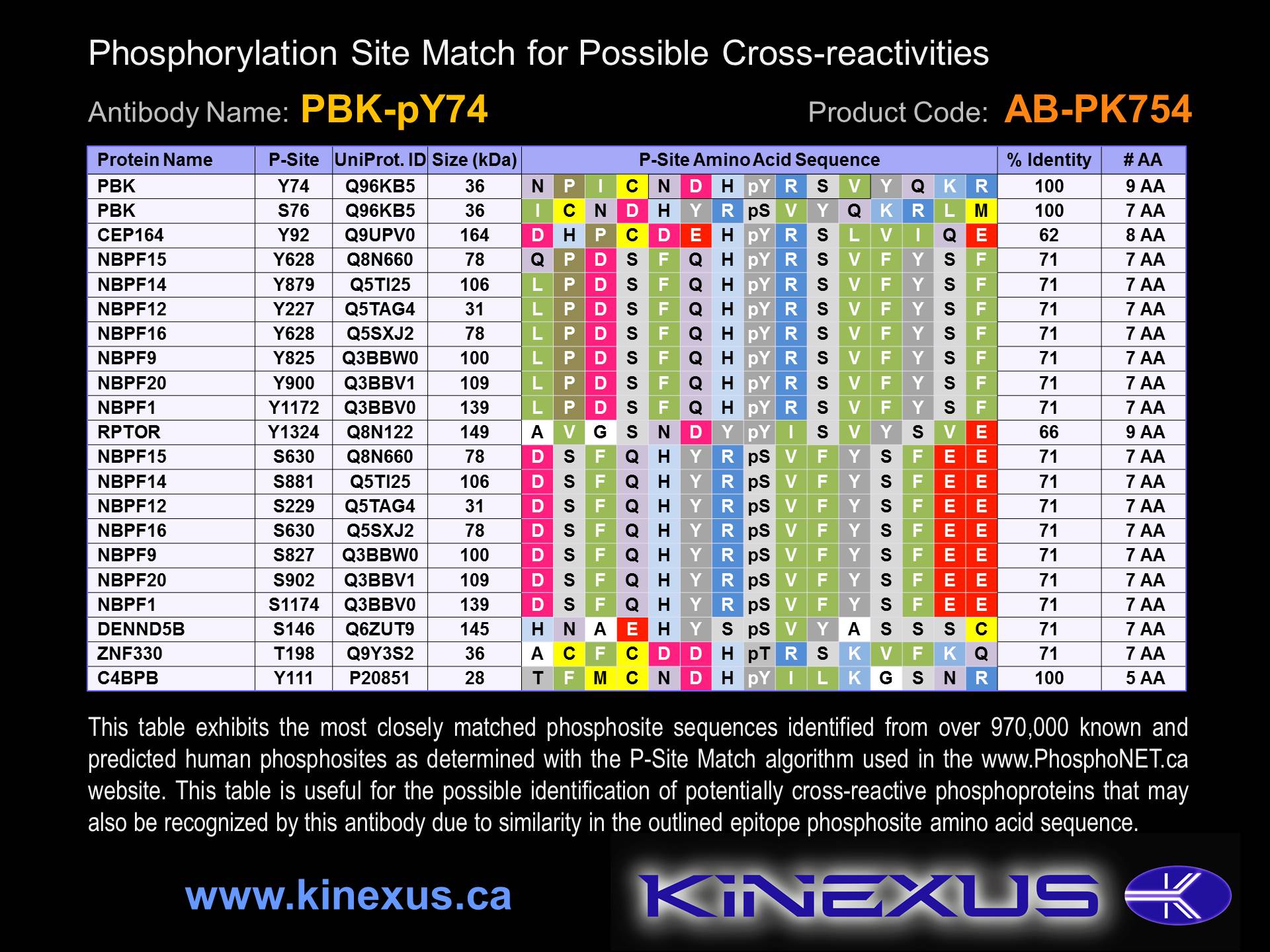
Figure 2. Identification of phosphosites related to PBK-pY74.

© Kinexus Bioinformatics Corporation 2017