
Product Name: PKCa-pT497
Product Number: AB-PK763
| Size: | 25 µg | Price: | 89.00 | |
| $US |
Target Full Name: Protein-serine kinase C alpha
Target Alias: AAG6; Kinase PKC-alpha; KPCA; PKC III; PKC-A; PKC-alpha; PKC-III; PRKACA; Protein kinase C, alpha; Protein kinase C, alpha type; MGC129900; MGC129901; CCDS11664.1; P17252; ENSG00000154229
Product Type Specific: Protein kinase phosphosite-specific antibody
Antibody Code: PK763
Antibody Target Type: Phosphosite-specific
Antibody Phosphosite: T497
Protein UniProt: P17252
Protein SigNET: P17252
Antibody Type: Polyclonal
Antibody Host Species: Rabbit
Antibody Immunogen Source: Human PKCa (PRKCA) sequence peptide Cat. No.: PE-04AOS99
Antibody Immunogen Sequence: TTR(pT)FCG(bA)C
Antibody Immunogen Description: Corresponds to amino acid residues T494 to G500; In the protein kinase catalytic domain activation T loop region between subdomains VII and VIII.
Antibody Immunogen Sequence: TTR(pT)FCG(bA)C
Antibody Immunogen Description: Corresponds to amino acid residues T494 to G500; In the protein kinase catalytic domain activation T loop region between subdomains VII and VIII.
Production Method: The immunizing peptide was produced by solid phase synthesis on a multipep peptide synthesizer and purified by reverse-phase hplc chromatography. Purity was assessed by analytical hplc and the amino acid sequence confirmed by mass spectrometry analysis. This peptide was coupled to KLH prior to immunization into rabbits. New Zealand White rabbits were subcutaneously injected with KLH-coupled immunizing peptide every 4 weeks for 4 months. The sera from these animals was applied onto an agarose column to which the immunogen peptide was thio-linked. Antibody was eluted from the column with 0.1 M glycine, pH 2.5. Subsequently, the antibody solution was neutralized to pH 7.0 with saturated Tris.This antibody was also subject to negative purification over phosphotyrosine-agarose.
Antibody Modification: Unconjugated. Contact KInexus if you are interest in having the antibody biotinylated or coupled with fluorescent dyes.
Antibody Modification: Unconjugated. Contact KInexus if you are interest in having the antibody biotinylated or coupled with fluorescent dyes.
Antibody Concentration: 1 mg/ml
Storage Buffer: Phosphate buffered saline pH 7.4, 0.05% Thimerasol
Storage Conditions: For long term storage, keep frozen at -40°C or lower. Stock solution can be kept at +4°C for more than 3 months. Avoid repeated freeze-thaw cycles.
Product Use: Western blotting | Antibody microarray
Antibody Dilution Recommended: 2 µg/ml for immunoblotting
Antibody Potency: Very strong immunoreactivity with immunogen peptide on dot blots.
Antibody Species Reactivity: Human
Antibody Positive Control: The observed molecular mass of the processed target protein on SDS-PAGE gels is reported to be around 75-90 kDa.
Related Product 1: PKCa-pT497 blocking peptide
Related Product 2: PKC-III (PKCa-1) pan-specific antibody (Cat. No.: AB-NK201)
Related Product 3: PKCa-pY195 phosphosite-specific antibody (Cat. No.: AB-PK764)
Related Product 4: CREB1 (123-135) KinSub - cyclic-AMP response element binding protein-1 (K123-R135, human) peptide; Crebtide protein kinase substrate peptide
Related Product 5: CREB1 (123-135) KinSub, biotinyl. - cyclic-AMP response element binding protein-1 (K123-R135, human) peptide; Crebtide protein kinase substrate peptide
Related Product 6: PKCA/BSubtide - PKCa (PRKCA) protein kinase substrate peptide
Related Product 7: PKCtide KinSub - Protein kinase C peptide substrate
Scientific Background: PKCa (PRKCA, PKC-alpha) is a protein-serine/threonine kinase in the classical protein kinase C family. This kinase is moderate to highly expressed in most tested human tissues. It is calcium activated, and dependent on acidic phospholipids (e.g. phosphatidylserine) and diacylglycerol for full phosphotransferase activity. Fatty acids can also activate PKCa. Phosphorylation of T494, T495 and T497 increases kinase activity. Phosphorylation of S657 increases phosphotransferase activity, protects against dephosphorylation of the T497 site and degradation of PKC-alpha. Phosphorylation of T638 is not essential for catalytic activity, but it protects against dephosphorylation of the T497 site. PKCa has been identified as a fundamental regulator of cardiac contractility and Ca(2+) handling in myocytes. PKCa has been linked with the development of colon, pituitary and thyroid cancers as well as glioblastoma multiforme (GM). PKCa has been considered as an oncoprotein (OP), since during tumourigenesis, as it is strongly activated by many different tumour promoting compounds, such as phorbol esters and teleocidins. While these compounds promote binding of PKC isoforms to the plasma membrane of cells, which stimulates their phosphotransferase activity, these agents also induce rapid down-regulation of PKC isoforms. Consequently, PKCa may be actually be a tumour suppressor protein. PKCa is highly expressed in several cancers, including malignant breast cancers and gliomas. Increased expression of PKCa has been observed in adenomatous pituitaries, with highest expression in invasive pituitary tumours. A point mutation was also identified in 4 invasive pituitary tumours. Furthermore, PKCa appears to stimulate cell proliferation, because many of its direct targets, such as Raf1, Bcl2, and CSPG4, participate in growth factor signalling pathways and PKC activators induce the downstream activation of ERK1/2 (MAPK3/1) and RAP1-GAP.
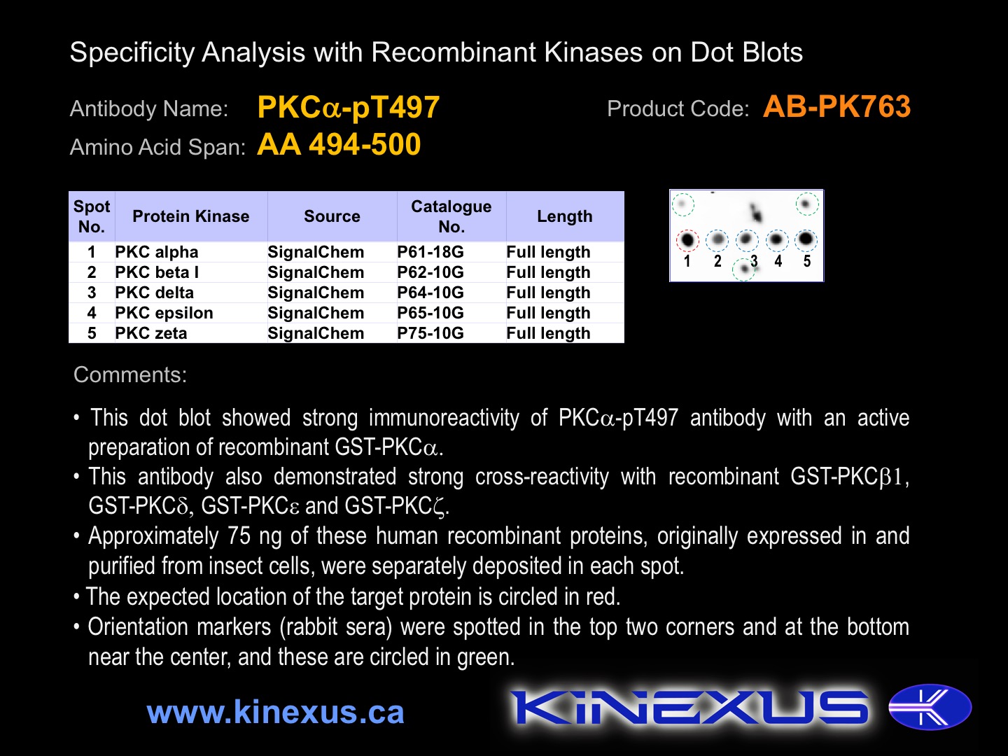
Figure 1. Dot blotting PKCa-pT497 antibody with recombinant purified proteins.
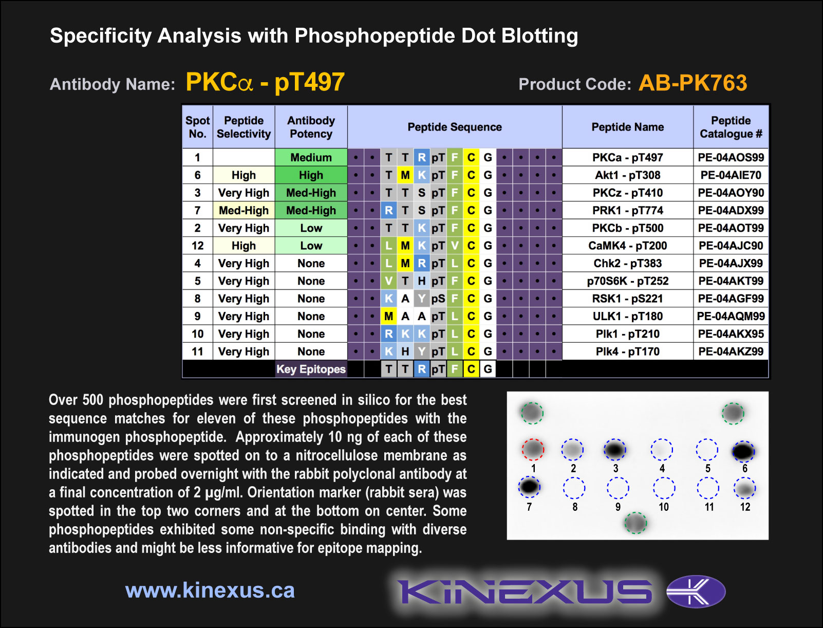
Figure 2. Epitope mapping of PKCa-pT497 antibody with similar phosphopeptides on dot blots.
Figure 3. Epitope mapping of PKCa-pT497 antibody with similar phosphopeptides on dot blots.

© Kinexus Bioinformatics Corporation 2017