
Product Name: PKCm-pS738+pS742
Product Number: AB-PK771
| Size: | 25 µg | Price: | 89.00 | |
| $US |
Target Full Name: Protein-serine kinase C mu (Protein kinase D)
Target Alias: Kinase PKD1; KPCD1; NPKC-mu; PKCM; PKC-mu; PKD; PRKCM; PRKD1; Protein kinase D1; Protein kinase C, mu type; Protein kinase D; ENSG00000184304
Product Type Specific: Protein kinase phosphosite-specific antibody
Antibody Code: PK771
Antibody Target Type: Phosphosite-specific
Antibody Phosphosite: S738+S742
Protein UniProt: Q15139
Protein SigNET: Q15139
Antibody Type: Polyclonal
Antibody Host Species: Rabbit
Antibody Immunogen Source: Human PKCm (PRKCM, PRKD1, PKD1) sequence peptide Cat. No.: PE-04AOZ95
Antibody Immunogen Sequence: GEK(pS)FRR(pS)VVG(bA)C
Antibody Immunogen Description: Corresponds to amino acid residues G735 to G745; In the protein kinase catalytic domain activation T loop region between subdomains VII and VIII.
Antibody Immunogen Sequence: GEK(pS)FRR(pS)VVG(bA)C
Antibody Immunogen Description: Corresponds to amino acid residues G735 to G745; In the protein kinase catalytic domain activation T loop region between subdomains VII and VIII.
Production Method: The immunizing peptide was produced by solid phase synthesis on a multipep peptide synthesizer and purified by reverse-phase hplc chromatography. Purity was assessed by analytical hplc and the amino acid sequence confirmed by mass spectrometry analysis. This peptide was coupled to KLH prior to immunization into rabbits. New Zealand White rabbits were subcutaneously injected with KLH-coupled immunizing peptide every 4 weeks for 4 months. The sera from these animals was applied onto an agarose column to which the immunogen peptide was thio-linked. Antibody was eluted from the column with 0.1 M glycine, pH 2.5. Subsequently, the antibody solution was neutralized to pH 7.0 with saturated Tris.This antibody was also subject to negative purification over phosphotyrosine-agarose.
Antibody Modification: Unconjugated. Contact KInexus if you are interest in having the antibody biotinylated or coupled with fluorescent dyes.
Antibody Modification: Unconjugated. Contact KInexus if you are interest in having the antibody biotinylated or coupled with fluorescent dyes.
Antibody Concentration: 0.25 mg/ml
Storage Buffer: Phosphate buffered saline pH 7.4, 0.05% Thimerasol
Storage Conditions: For long term storage, keep frozen at -40°C or lower. Stock solution can be kept at +4°C for more than 3 months. Avoid repeated freeze-thaw cycles.
Product Use: Western blotting | Antibody microarray
Antibody Dilution Recommended: 2 µg/ml for immunoblotting
Antibody Species Reactivity: Human
Antibody Positive Control: The observed molecular mass of the processed target protein on SDS-PAGE gels is reported to be around 100-125 kDa.
Antibody Specificity: Very high
Antibody Cross Reactivity: No significant cross-reactive proteins detected in phenylarsine oxide (PAO)+vanadate-treated HeLa cells, EGF-treated A431 cells and insulin-treated MCF7 cells, when these cells were homogenized in SDS-PAGE sample buffer.
Related Product 1: PKCm-pS738+pS742 blocking peptide
Related Product 2: PKCm-pS205 phosphosite-specific antibody (Cat. No.: AB-PK770)
Related Product 3: PKCMSubtide - PKD1 (PRKCM) protein kinase substrate peptide
Related Product 1: PKCm-pS738+pS742 blocking peptide
Related Product 2: PKCm-pS205 phosphosite-specific antibody (Cat. No.: AB-PK770)
Related Product 3: PKCMSubtide - PKD1 (PRKCM) protein kinase substrate peptide
Scientific Background: PKD1 (PRKD1, PKCm, PKC-mu) is a protein-serine/threonine kinase of the CAMK group and PKD family. It is a protein in the novel protein kinase C family. It is moderate to highly expressed in most tested human tissues, especially in the thymus, lung and peripheral blood mononuclear cells, but poorly expressed in the brain and spinal cord. It is dependent on acidic phospholipids (e.g. phosphatidylserine) and diacylglycerol (DAG) for full phosphotransferase activity, and does not require calcium. PKD1 is activated by DAG and phorbol esters, which via its phorbol-ester/DAG-type domain 1 binds DAG with high affinity and appears to play the dominant role in mediating translocation to the cell membrane and trans-Golgi network. Phorbol-ester/DAG-type domain 2 binds phorbol ester with higher affinity. Phosphorylation at Y95 increases kinase activity and induces interaction with PKC-delta. Phosphorylation at S249, Y463, S738, S742 and S910 increases kinase activity. Autophosphorylation of S742 and phosphorylation of S738 by PKC relieves auto-inhibition by the PH domain, and also induces interaction with ASK1, JNK1 and IKKb. Phosphorylation at S205, S208, S219 and S223 induces binding of 14-3-3 beta and tau to inhibit PKD1 phosphotransferase activity. Phosphorylation at S397 induces interaction with 14-3-3 beta. PKD1 forms a complex in vivo with a PI4-kinase and a PI4-phosphate 5-kinase. A region of PKD1 between the amino-terminal transmembrane domain and the pleckstrin homology domain has been shown to be involved in the association with the lipid kinases . PKD1 is thought to have a role in regulating cellular trafficking, actin remodeling, gene transcription and protection from oxidative stress. PKD1 is implicated in prostate cancer through phosphorylation of e-cadherin leading to increased malignancy and motility of tumours. PKD1 has also been linked with the development of colorectal adenocarcinomas, lung bronchoalveolar carcinomas and melanomas (metastatic).
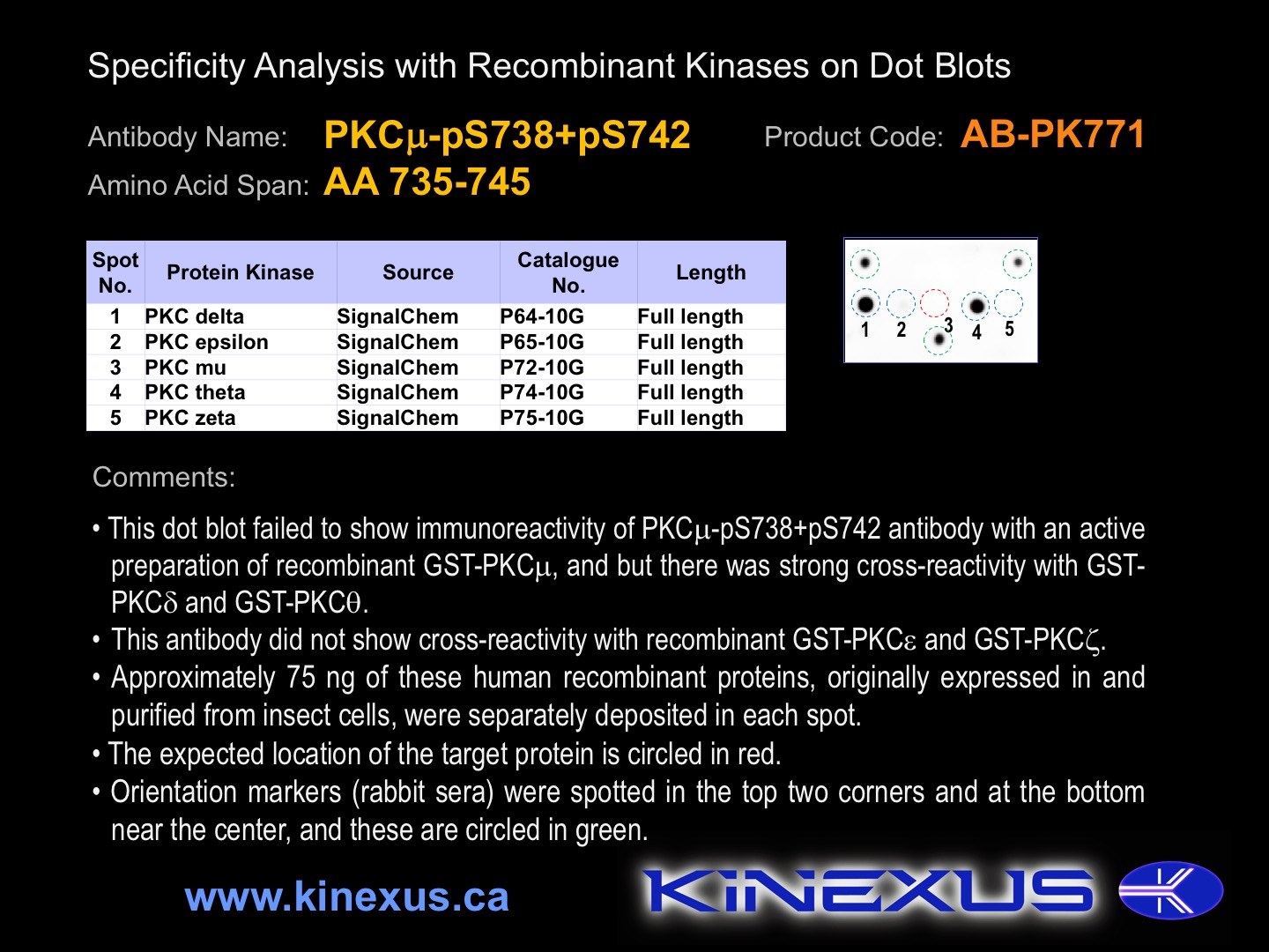
Figure 1. Dot blotting PKCm-pS738+pS742 antibody with recombinant purified proteins.
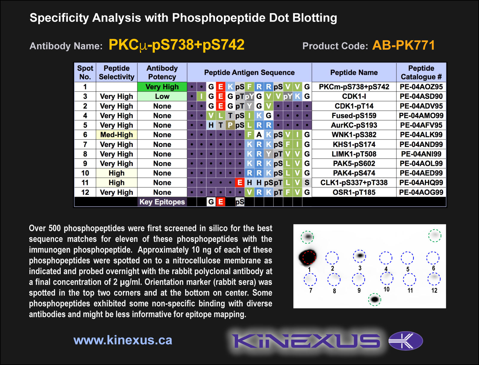
Figure 2. Epitope mapping of PKCm-pS738+pS742 antibody with similar phosphopeptides on dot blots.
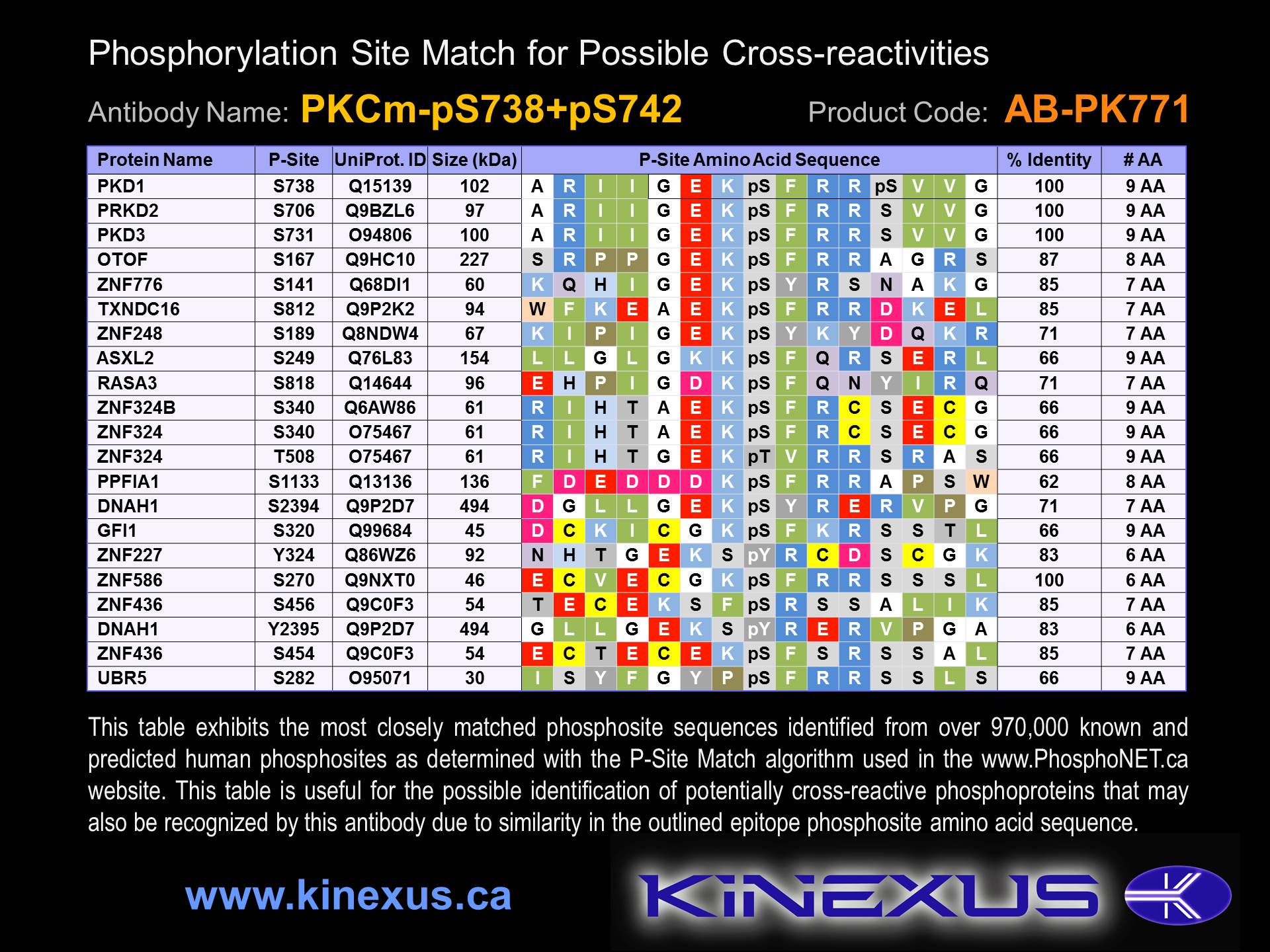
Figure 3. Identification of phosphosites related to PKCm-pS738+pS742.

© Kinexus Bioinformatics Corporation 2017