
Product Name: PKCq-pS695
Product Number: AB-PK772
| Size: | 25 µg | Price: | 89.00 | |
| $US |
Target Full Name: Protein-serine kinase C theta
Target Alias: Kinase PKC-theta; KPCT; MGC126514; MGC141919; NPKC-theta; PKCQ; PKC-theta; PRKCQ; PRKCT; Protein kinase C, theta; Protein kinase C, theta type; CCDS7079.1; Q04759; ENSG00000065675
Product Type Specific: Protein kinase phosphosite-specific antibody
Antibody Code: PK772
Antibody Target Type: Phosphosite-specific
Antibody Phosphosite: S695
Protein UniProt: Q04759
Protein SigNET: Q04759
Antibody Type: Polyclonal
Antibody Host Species: Rabbit
Antibody Immunogen Source: Human PKCt (PRKCQ) sequence peptide Cat. No.: PE-04AOX99
Antibody Immunogen Sequence: RNF(pS)FMN(bA)C
Antibody Immunogen Description: Corresponds to amino acid residues R692 to N698; In Pkinase_C domain near C-terminus of the kinase
Antibody Immunogen Sequence: RNF(pS)FMN(bA)C
Antibody Immunogen Description: Corresponds to amino acid residues R692 to N698; In Pkinase_C domain near C-terminus of the kinase
Production Method: The immunizing peptide was produced by solid phase synthesis on a multipep peptide synthesizer and purified by reverse-phase hplc chromatography. Purity was assessed by analytical hplc and the amino acid sequence confirmed by mass spectrometry analysis. This peptide was coupled to KLH prior to immunization into rabbits. New Zealand White rabbits were subcutaneously injected with KLH-coupled immunizing peptide every 4 weeks for 4 months. The sera from these animals was applied onto an agarose column to which the immunogen peptide was thio-linked. Antibody was eluted from the column with 0.1 M glycine, pH 2.5. Subsequently, the antibody solution was neutralized to pH 7.0 with saturated Tris.This antibody was also subject to negative purification over phosphotyrosine-agarose.
Antibody Modification: Unconjugated. Contact KInexus if you are interest in having the antibody biotinylated or coupled with fluorescent dyes.
Antibody Modification: Unconjugated. Contact KInexus if you are interest in having the antibody biotinylated or coupled with fluorescent dyes.
Antibody Concentration: 1 mg/ml
Storage Buffer: Phosphate buffered saline pH 7.4, 0.05% Thimerasol
Storage Conditions: For long term storage, keep frozen at -40°C or lower. Stock solution can be kept at +4°C for more than 3 months. Avoid repeated freeze-thaw cycles.
Product Use: Western blotting | Antibody microarray
Antibody Dilution Recommended: 2 µg/ml for immunoblotting
Antibody Potency: Strong immunoreactivity of a target-sized protein by Western blotting in Jurkat cells. Very strong immunoreactivity with immunogen peptide on dot blots.
Antibody Species Reactivity: Human
Antibody Positive Control: The observed molecular mass of the processed target protein on SDS-PAGE gels is reported to be around 75-84 kDa.
Antibody Specificity: Very high
Antibody Cross Reactivity: No significant cross-reactive proteins detected in T98G cells.
Related Product 1: PKCq-pS695 blocking peptide
Related Product 2: PKCq-pY545 phosphosite-specific antibody (Cat. No.: AB-PK773)
Antibody Specificity: Very high
Antibody Cross Reactivity: No significant cross-reactive proteins detected in T98G cells.
Related Product 1: PKCq-pS695 blocking peptide
Related Product 2: PKCq-pY545 phosphosite-specific antibody (Cat. No.: AB-PK773)
Scientific Background: PKCt (PKCq, PRKCT, PKC-theta) is a protein-serine/threonine kinase of the AGC group and novel PKC family. It is dependent on acidic phospholipids (e.g. phosphatidylserine) and diacylglycerol for full phosphotransferase activity, and does not require calcium. It is essential in TCR signalling for T-cell activation, differentiation, proliferation, and survival. These T-cell responses are mediated via Jun, NF-kappa-B, NFATC1 and NFATC2. The phosphotransferase activity of PKCt is unaffected by an Y90F mutation, but T-cell proliferation is inhibited. An A148E mutation results in constitutive activation of PKCt. A T219A mutation again will not inhibit kinase phosphotransferase activity, but it will inhibit localization to the plasma membrane and inhibit transactivation of IL2 promoter. Phosphorylation at Y90, T538 (activation loop of the kinase domain) and S676 (turn motif) increases phosphotransferase activity. Phosphorylation at S695 (hydrophobic region) increases phosphotransferase activity and interaction with PDK1. Insertional mutagenesis studies in mice support a role for this protein kinase in mouse cancer oncogenesis. PKCt is linked to Gastrointestinal Stromal Tumours (GANT, GIST), which are rare sarcomas arising from tissues lining the gastrointestinal tract and typically arise from the stomach, intestines, or colon. GANT has been related to neurofibromatosis and adenocarcinoma as well.
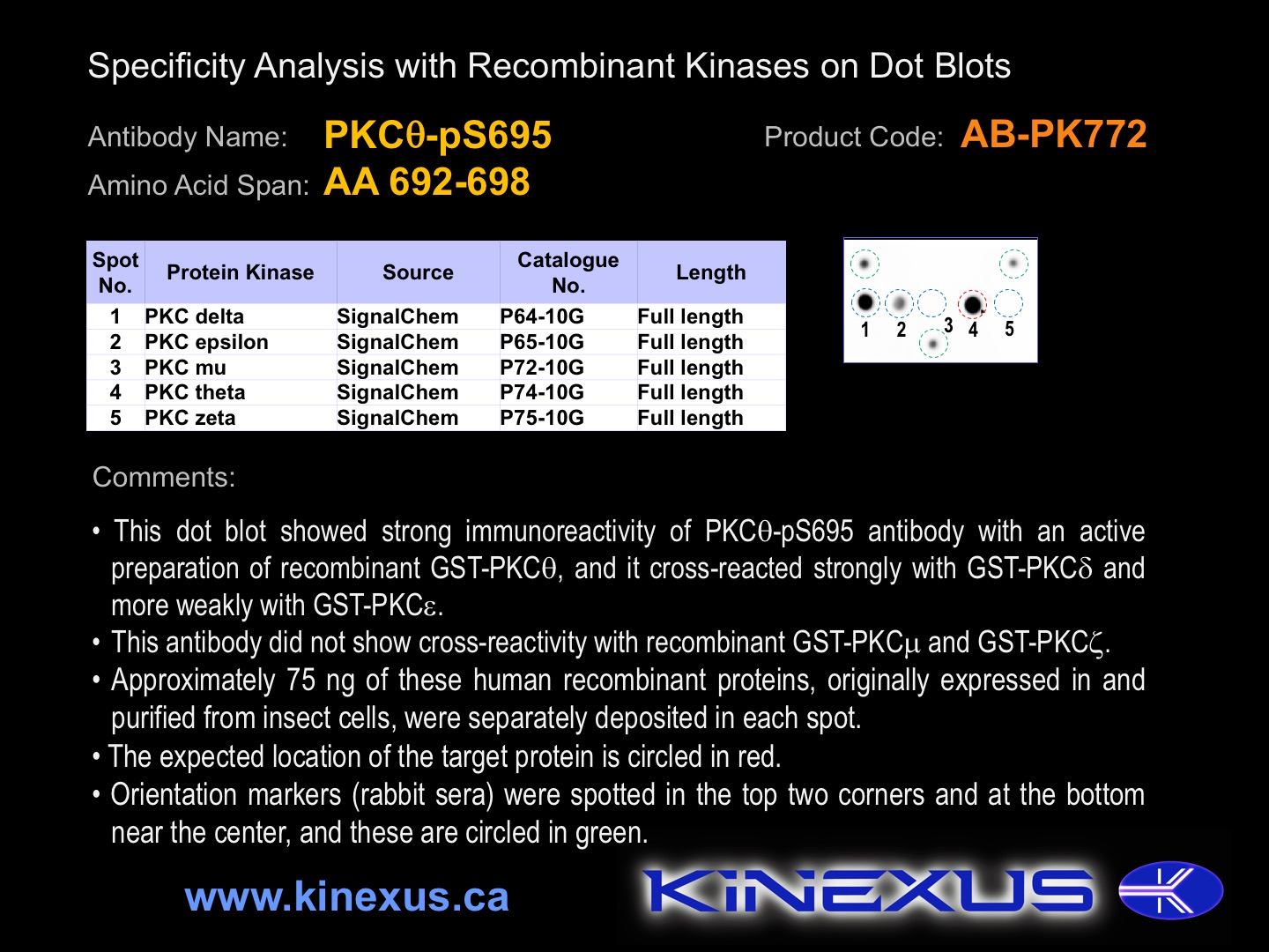
Figure 1. Dot blotting PKCq-pS695 antibody with recombinant purified proteins.
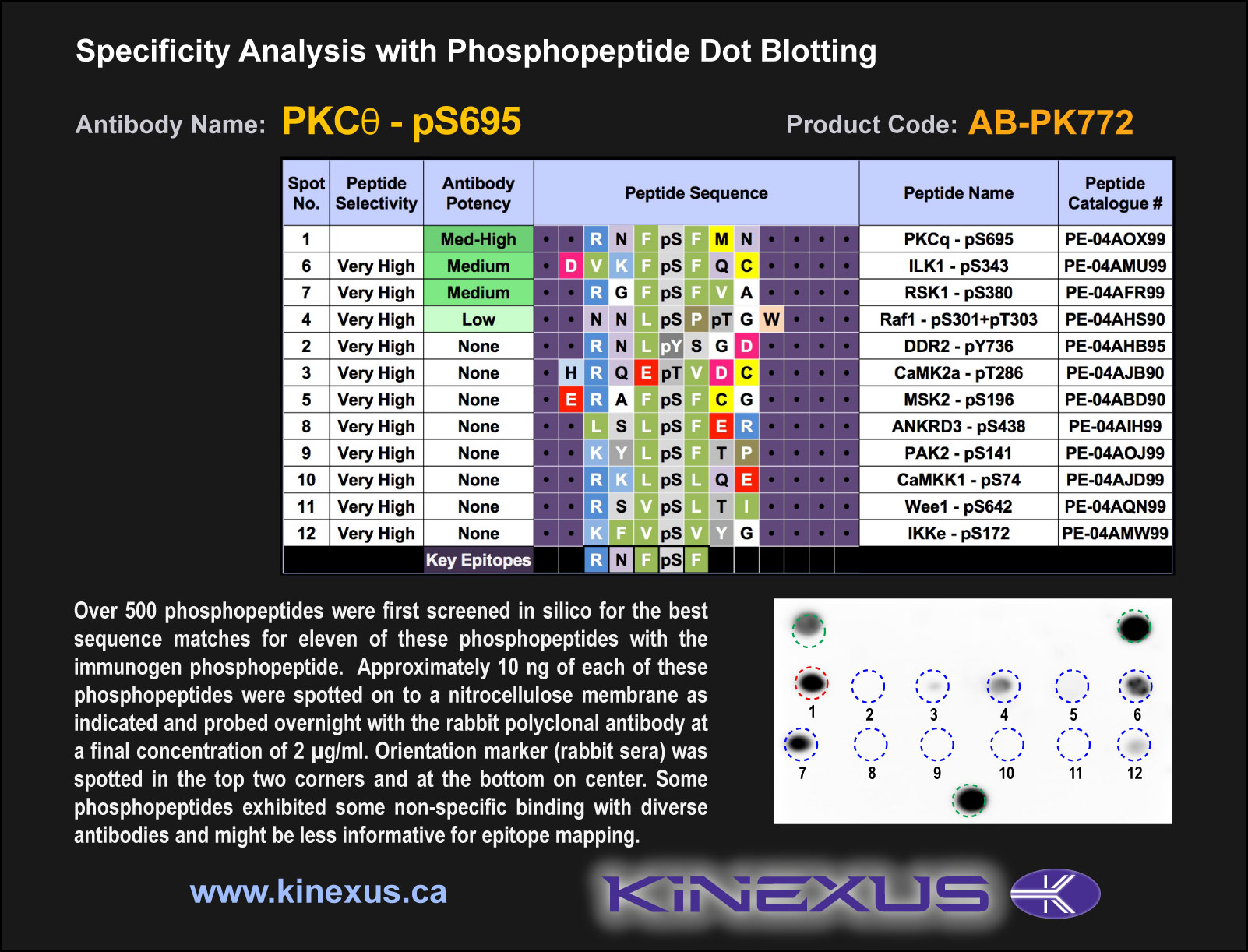
Figure 2. Epitope mapping of PKCq-pS695 antibody with similar phosphopeptides on dot blots.
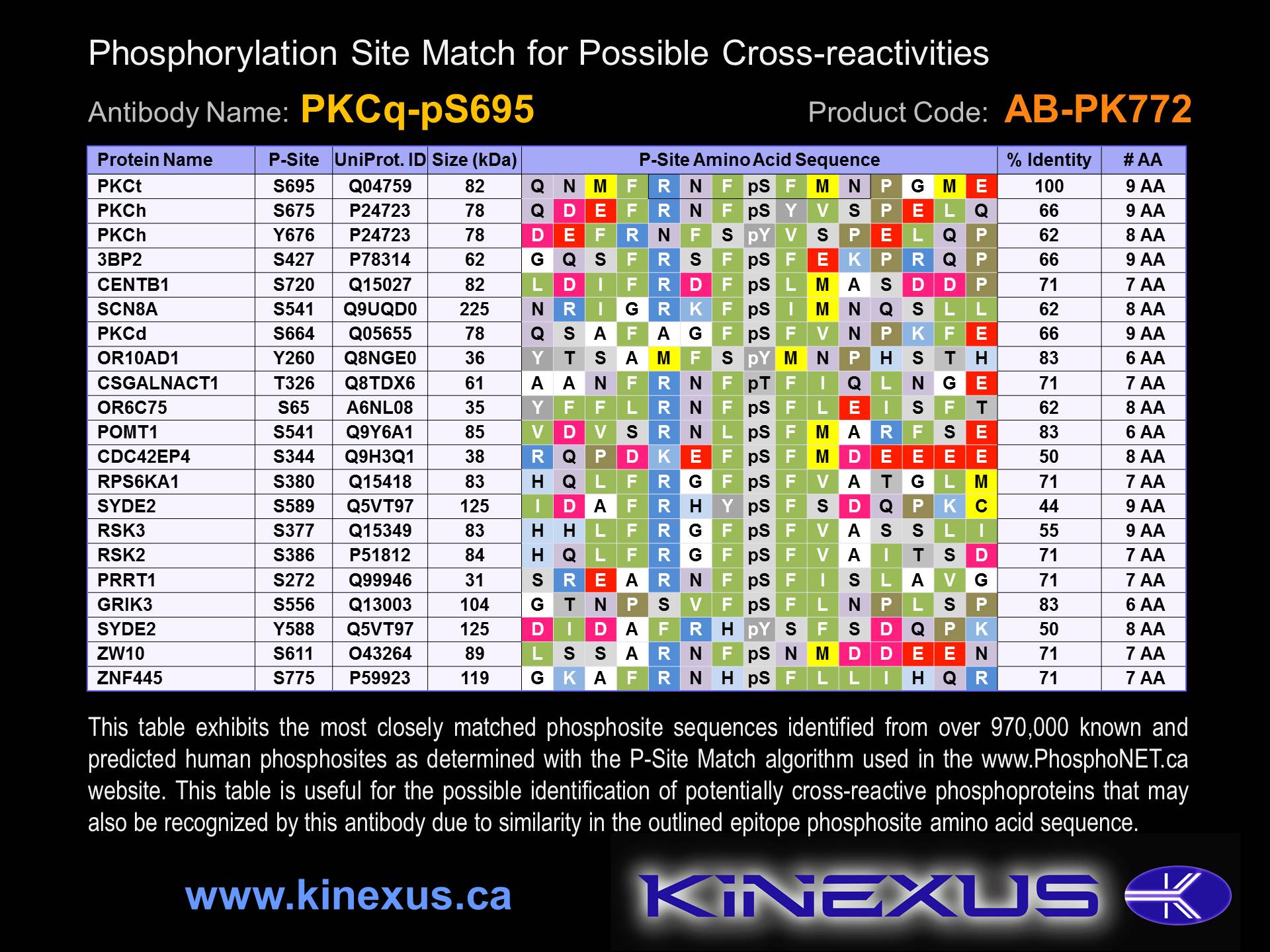
Figure 3. Identification of phosphosites related to PKCq-pS695.

© Kinexus Bioinformatics Corporation 2017