
Product Name: TAOK1-pY309
Product Number: AB-PK827
| Size: | 25 µg | Price: | 89.00 | |
| $US |
Target Full Name: Serine/threonine-protein kinase TAO1
Target Alias: FLJ14314; HKFC-B; KIAA1361; MAP3K16; MARKK; PSK2; STE20-like kinase PSK2; TAOK1; TAO kinase 1; KFC-B; MARKK; h KFC-B; ENSG00000160551
Product Type Specific: Protein kinase phosphosite-specific antibody
Antibody Code: PK827
Antibody Target Type: Phosphosite-specific
Antibody Phosphosite: Y309
Protein UniProt: Q7L7X3
Protein SigNET: Q7L7X3
Antibody Type: Polyclonal
Antibody Host Species: Rabbit
Antibody Immunogen Source: Human TAO1 (TAOK1) sequence peptide Cat. No.: PE-04APX95
Antibody Immunogen Sequence: NLQ(pY)RKM(bA)C
Antibody Immunogen Description: Corresponds to amino acid residues N306 to M312; Just after the kinase catalytic domain. Thhis is the major in vivo site of phosphorylation on TAO1.
Antibody Immunogen Sequence: NLQ(pY)RKM(bA)C
Antibody Immunogen Description: Corresponds to amino acid residues N306 to M312; Just after the kinase catalytic domain. Thhis is the major in vivo site of phosphorylation on TAO1.
Production Method: The immunizing peptide was produced by solid phase synthesis on a multipep peptide synthesizer and purified by reverse-phase hplc chromatography. Purity was assessed by analytical hplc and the amino acid sequence confirmed by mass spectrometry analysis. This peptide was coupled to KLH prior to immunization into rabbits. New Zealand White rabbits were subcutaneously injected with KLH-coupled immunizing peptide every 4 weeks for 4 months. The sera from these animals was applied onto an agarose column to which the immunogen peptide was thio-linked. Antibody was eluted from the column with 0.1 M glycine, pH 2.5. Subsequently, the antibody solution was neutralized to pH 7.0 with saturated Tris.This antibody was also subject to negative purification over phosphotyrosine-agarose.
Antibody Modification: Unconjugated. Contact KInexus if you are interest in having the antibody biotinylated or coupled with fluorescent dyes.
Antibody Modification: Unconjugated. Contact KInexus if you are interest in having the antibody biotinylated or coupled with fluorescent dyes.
Antibody Concentration: 1 mg/ml
Storage Buffer: Phosphate buffered saline pH 7.4, 0.05% Thimerasol
Storage Conditions: For long term storage, keep frozen at -40°C or lower. Stock solution can be kept at +4°C for more than 3 months. Avoid repeated freeze-thaw cycles.
Product Use: Western blotting | Antibody microarray
Antibody Dilution Recommended: 2 µg/ml for immunoblotting
Antibody Potency: Strong immunoreactivity of a target-sized protein by Western blotting in HeLa cells and weak detection in HepG2 cells. Very strong immunoreactivity with immunogen peptide on dot blots.
Antibody Species Reactivity: Human
Antibody Positive Control: The observed molecular mass of the processed target protein on SDS-PAGE gels is reported to be around 110-125 kDa.
Antibody Specificity: High
Antibody Cross Reactivity: No significant cross-reactive proteins detected in HepG2, MCF7 and T98G cells, except for ~80 kDa protein in HeLa cells and ~65 kDa protein in inulin-treated MCF7 cells.
Related Product 1: TAO1-pY309 blocking peptide
Related Product 2: TAO1-pS181 phosphosite-specific antibody (Cat. No.: AB-PK826)
Related Product 3: TAOSubtide - TAOK2 (PSK) protein kinase substrate peptide
Scientific Background: TAO1 (MAP3K16, TAOK1) is a protein-serine/threonine kinase of the STE group and STE20 family. It is involved in various processes such as p38/MAPK14 stress-activated MAPK cascade, DNA damage response and regulation of cytoskeleton stability. It phosphorylates MAP2K3, MAP2K6 and MARK2, and acts as an activator of the p38 (MAPK14) stress-activated MAPK cascade by phosphorylation and activation of the upstream MKK3 and MKK6 kinases. It is involved in G protein-coupled receptor signalling to p38 (MAPK14). In response to DNA damage, it is involved in the G2/M transition DNA damage checkpoint by activating the p38 (MAPK14) stress-activated MAPK cascade, probably by mediating phosphorylation of MKK3 (MAP2K3) and MKK6 (MAP2K6). It serves as a regulator of cytoskeleton stability by phosphorylating MARK2 at T208, leading to its activation and subsequent phosphorylation and detachment of MAPT/TAU from microtubules. It also regulates apoptotic morphological changes, including cell contraction, membrane blebbing and apoptotic bodies formation via activation of the JNK (MAPK8) cascade.
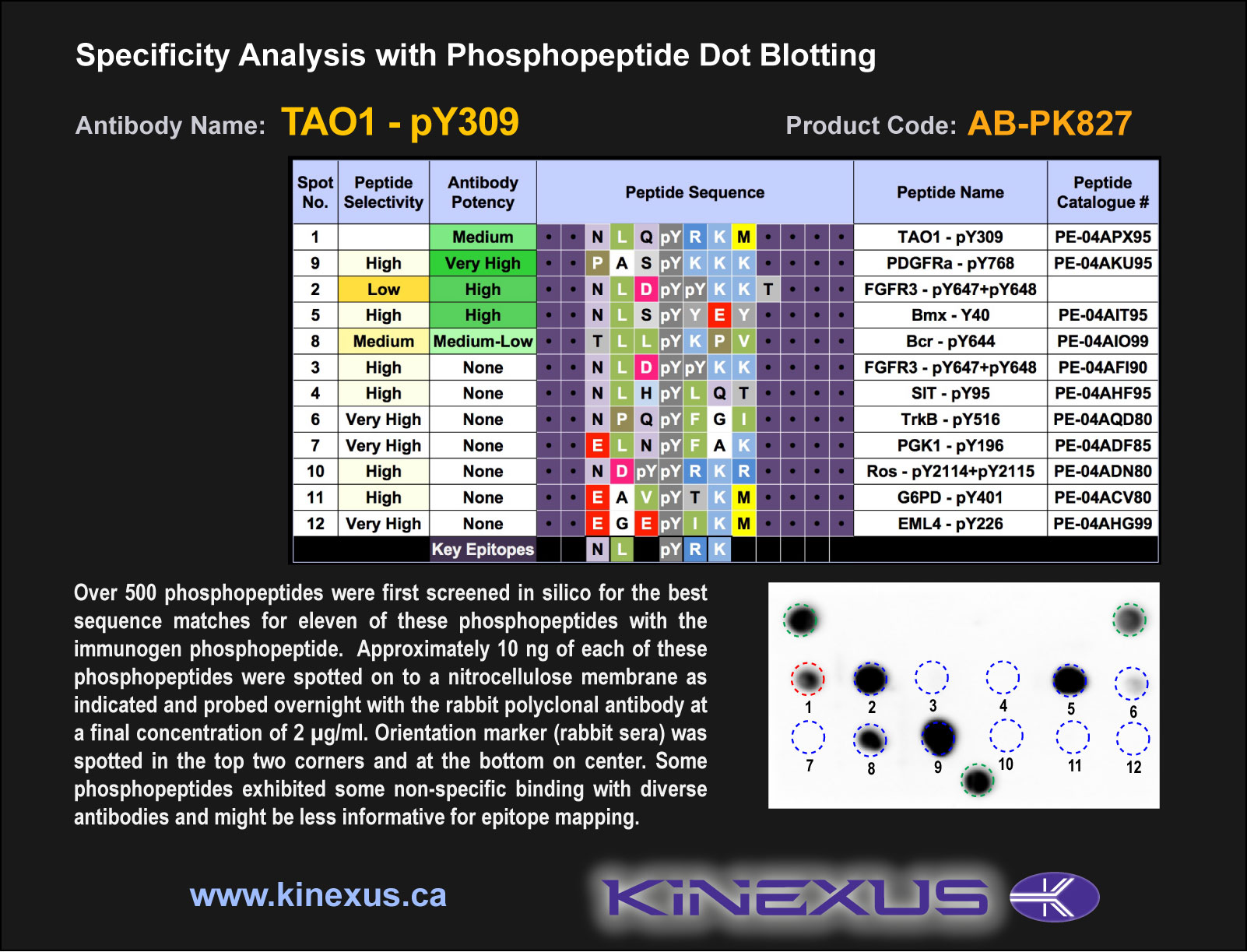
Figure 1. Epitope mapping of TAO1-pY309 antibody with similar phosphopeptides on dot blots.
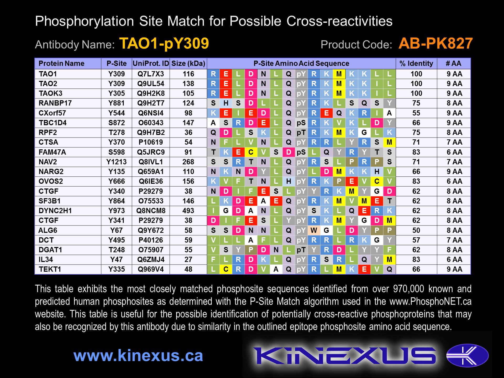
Figure 2. Identification of phosphosites related to TAO1-pY309.

© Kinexus Bioinformatics Corporation 2017