
Product Name: Wee1-pS642
Product Number: AB-PK854
| Size: | 25 µg | Price: | 89.00 | |
| $US |
Target Full Name: Wee1 protein-tyrosine kinase
Target Alias: Kinase WEE1; WEE1; WEE1A; WEE1hu
Product Type Specific: Protein kinase phosphosite-specific antibody
Antibody Code: PK854
Antibody Target Type: Phosphosite-specific
Antibody Phosphosite: S642
Protein UniProt: P30291
Protein SigNET: P30291
Antibody Type: Polyclonal
Antibody Host Species: Rabbit
Antibody Immunogen Source: Human Wee1 sequence peptide Cat. No.: PE-04AQN99
Antibody Immunogen Sequence: RSV(pS)LTI(bA)C
Antibody Immunogen Description: Corresponds to amino acid residues R639 to I645; In the C-terminus of the kinase
Antibody Immunogen Description: Corresponds to amino acid residues R639 to I645; In the C-terminus of the kinase
Production Method: The immunizing peptide was produced by solid phase synthesis on a multipep peptide synthesizer and purified by reverse-phase hplc chromatography. Purity was assessed by analytical hplc and the amino acid sequence confirmed by mass spectrometry analysis. This peptide was coupled to KLH prior to immunization into rabbits. New Zealand White rabbits were subcutaneously injected with KLH-coupled immunizing peptide every 4 weeks for 4 months. The sera from these animals was applied onto an agarose column to which the immunogen peptide was thio-linked. Antibody was eluted from the column with 0.1 M glycine, pH 2.5. Subsequently, the antibody solution was neutralized to pH 7.0 with saturated Tris.This antibody was also subject to negative purification over phosphotyrosine-agarose.
Antibody Modification: Unconjugated. Contact KInexus if you are interest in having the antibody biotinylated or coupled with fluorescent dyes.
Antibody Modification: Unconjugated. Contact KInexus if you are interest in having the antibody biotinylated or coupled with fluorescent dyes.
Antibody Concentration: 1 mg/ml
Storage Buffer: Phosphate buffered saline pH 7.4, 0.05% Thimerasol
Storage Conditions: For long term storage, keep frozen at -40°C or lower. Stock solution can be kept at +4°C for more than 3 months. Avoid repeated freeze-thaw cycles.
Product Use: Western blotting | Antibody microarray
Antibody Dilution Recommended: 2 µg/ml for immunoblotting
Antibody Potency: Strong immunoreactivity of a target-sized protein by Western blotting in sea star oocytes. Very weak detection of target in MCF7 and T98G cells. Strong immunoreactivity with immunogen peptide on dot blots.
Antibody Species Reactivity: Human
Antibody Positive Control: The observed molecular mass of the processed target protein on SDS-PAGE gels is reported to be around 70-74 kDa.
Antibody Specificity: High
Antibody Cross Reactivity: Almost no significant cross-reactivities detected in HeLa cells, except for a 25 kDa protein; phenylarsine oxide (PAO) increases strong 50 kDa cross-reactive protein in HeLa cells. The 25 kDa cross-reactive protein was also detected in sea star oocytes and MCF7 and T98G cells.
Related Product 1: Wee1-pS642 blocking peptide
Scientific Background: Wee1 is a Dual specificity protein kinase of the Other group and WEE family. It is a negative regulator to entry into G2-M transition in mitosis through phosphorylation of CDK1. Wee1 is a nuclear protein that catalyzes the inhibitory tyrosine phosphorylation of CDC2/cyclin B kinase, and appears to coordinate the transition between DNA replication and mitosis by protecting the nucleus from cytoplasmically activated CDC2 kinase. Phosphorylatio of S121 induces proteolysis through interaction with BTRC and FBW1B. Phosphorylation of S53 and S123 beta-transducin repeat-containing protein recognition for degradation by ubiquitination. Phosphorylation of T187, T190 and S193 induces proteolysis through interaction with BTRC and FBW1B. Phosphorylation of S642 inhibits phosphotransferase activity and induces interaction with 14-3-3 theta. Wee1 is highly expressed in testis. Wee1 kinase phosphorylates the p34 (CDC2) -cyclin B complex on Y15 and inactivates the p34 (CDC2)-cyclin B kinase. Wee1 is a target for cancer therapy. Specific inhibition of Wee1 can be used to increase the sensitivity of T-cell leukemia and pancreatic cancer. Wee1 was found to be a novel independent prognostic marker of poor survival ovarian carcinomas.
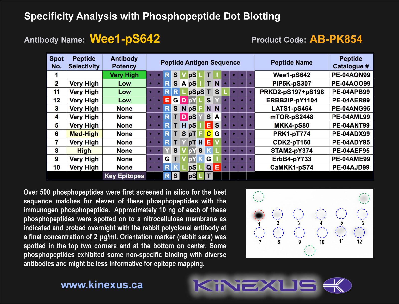
Figure 1. Epitope mapping of Wee1-pS642 antibody with similar phosphopeptides on dot blots.
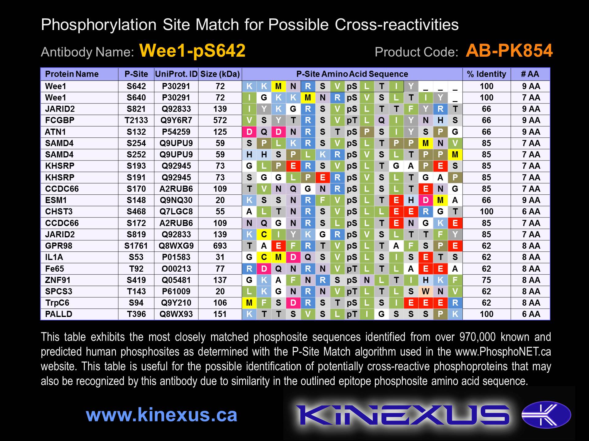
Figure 2. Identification of phosphosites related to Wee1-pS642.

© Kinexus Bioinformatics Corporation 2017