
Product Name: WNK1-pT60
Product Number: AB-PK856
| Size: | 25 µg | Price: | 89.00 | |
| $US |
Target Full Name: Serine/threonine-protein kinase WNK1
Target Alias: HSAN2; HSN2; KDP; KIAA0344; MGC163339; MGC163341; p65; PRKWNK1; Protein kinase with no lysine 1; Protein kinase, lysine-deficient 1; PSK; PHA2C; CCDS8506.1; ENSG00000060237
Product Type Specific: Protein kinase phosphosite-specific antibody
Antibody Code: PK856
Antibody Target Type: Phosphosite-specific
Antibody Phosphosite: T60
Protein UniProt: Q9H4A3
Protein SigNET: Q9H4A3
Antibody Type: Polyclonal
Antibody Host Species: Rabbit
Antibody Immunogen Source: Human WNK1 sequence peptide Cat. No.: PE-04ALJ99
Antibody Immunogen Sequence: RRH(pT)MDK(bA)C
Antibody Immunogen Description: Corresponds to amino acid residues R57 to K63; In the N-terminal portion of the protein kinase
Antibody Immunogen Sequence: RRH(pT)MDK(bA)C
Antibody Immunogen Description: Corresponds to amino acid residues R57 to K63; In the N-terminal portion of the protein kinase
Production Method: The immunizing peptide was produced by solid phase synthesis on a multipep peptide synthesizer and purified by reverse-phase hplc chromatography. Purity was assessed by analytical hplc and the amino acid sequence confirmed by mass spectrometry analysis. This peptide was coupled to KLH prior to immunization into rabbits. New Zealand White rabbits were subcutaneously injected with KLH-coupled immunizing peptide every 4 weeks for 4 months. The sera from these animals was applied onto an agarose column to which the immunogen peptide was thio-linked. Antibody was eluted from the column with 0.1 M glycine, pH 2.5. Subsequently, the antibody solution was neutralized to pH 7.0 with saturated Tris.This antibody was also subject to negative purification over phosphotyrosine-agarose.
Antibody Modification: Unconjugated. Contact KInexus if you are interest in having the antibody biotinylated or coupled with fluorescent dyes.
Antibody Modification: Unconjugated. Contact KInexus if you are interest in having the antibody biotinylated or coupled with fluorescent dyes.
Antibody Concentration: 1 mg/ml
Storage Buffer: Phosphate buffered saline pH 7.4, 0.05% Thimerasol
Storage Conditions: For long term storage, keep frozen at -40°C or lower. Stock solution can be kept at +4°C for more than 3 months. Avoid repeated freeze-thaw cycles.
Product Use: Western blotting | Antibody microarray
Antibody Dilution Recommended: 2 µg/ml for immunoblotting
Antibody Potency: Strong immunoreactivity with immunogen peptide on dot blots.
Antibody Species Reactivity: Human
Antibody Positive Control: The observed molecular mass of the processed target protein on SDS-PAGE gels is reported to be around 190, 250-400 kDa.
Antibody Specificity: Very high
Antibody Cross Reactivity: Almost no significant cross-reactivities detected in A431 and Jurkat cells, except for a 75 kDa cross-reactive protein that is also detected in sea star oocytes. Sea star oocytes also feature 140 and 200 kDa strongly cross-reactive proteins.
Related Product 1: WNK1-pT60 blocking peptide
Related Product 2: WNK1-1 pan-specific antibody (Cat. No.: AB-NK252-1)
Related Product 3: WNK1-2 pan-specific antibody (Cat. No.: AB-NK252-2)
Related Product 4: WNK1-3 pan-specific antibody (Cat. No.: AB-NK252-3)
Related Product 5: WNK1-pS382 phosphosite-specific antibody (Cat. No.: AB-PK855)
Scientific Background: WNK1 (PRKWNK1) is a protein-serine/threonine kinase of the Other group and WNK family. It is involved in electrolyte homeostasis, cell survival, cell signalling, and cell proliferation. It is activated by hypertonicity. The activation requires autophosphorylation of S382. Phosphorylation of S378 also promotes increased phosphotransferase activity. WNK1 can activate sodium-coupled chloride cotransporters, and inhibit potassium-coupled chloride cotransporters. Proteins activated by WNK1 include SCNN1A, SCNN1B, SCNN1D and SGK1 while an inhibited protein is WNK4 (via phosphorylation of WNK4 or an associated complex of a WNK1 autoinhibitory domain bound to WNK4). WNK4 inhibition could modulate, via phosphorylation, a thiazide-sensitive NaCl cotransporter transporter (specifically SLC12A3). WNK1 has also been proposed to modulate NEDD4L phosphorylation, and have an effect on actin cytoskeleton rearrangement. Insertional mutagenesis studies in mice support a role for this protein kinase in mouse cancer oncogenesis.

Figure 1. Epitope mapping of WNK1-pT60 antibody with similar phosphopeptides on dot blots.
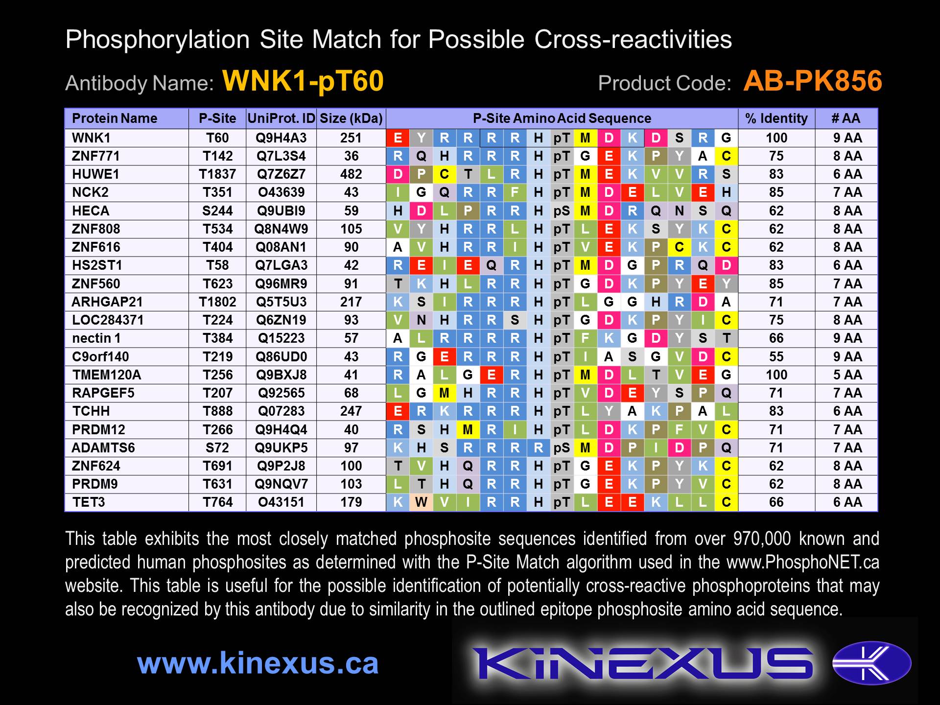
Figure 2. Identification of phosphosites related to WNK1-pT60.

© Kinexus Bioinformatics Corporation 2017