
Product Name: ERK1-pY204
Product Number: AB-PK864
| Size: | 25 µg | Price: | 89.00 | |
| $US |
Target Full Name: Extracellular regulated protein-serine kinase 1 (p44 MAP kinase)
Target Alias: ERK-1; ERT2; Insulin-stimulated MAP2 kinase; Kinase ERK1; MAP kinase 1; MAPK 1; MAPK1; MAPK3; PRKM3; p44ERK1; p44MAPK; MGC20180; ENSG00000102882
Product Type Specific: Protein kinase phosphosite-specific antibody
Antibody Code: PK864
Antibody Target Type: Phosphosite-specific
Antibody Phosphosite: Y204
Protein UniProt: P27361
Protein SigNET: P27361
Antibody Type: Polyclonal
Antibody Host Species: Rabbit
Antibody Immunogen Source: Human ERK1 (MAPK3) sequence peptide Cat. No.: PE-04ATK95
Antibody Immunogen Sequence: C(bA)(pY)VATRW
Antibody Immunogen Description: Corresponds to amino acid residues Y204 to W209; In activation T-loop between kinase catalytic subdomains VII and VIII
Antibody Immunogen Sequence: C(bA)(pY)VATRW
Antibody Immunogen Description: Corresponds to amino acid residues Y204 to W209; In activation T-loop between kinase catalytic subdomains VII and VIII
Production Method: The immunizing peptide was produced by solid phase synthesis on a multipep peptide synthesizer and purified by reverse-phase hplc chromatography. Purity was assessed by analytical hplc and the amino acid sequence confirmed by mass spectrometry analysis. This peptide was coupled to KLH prior to immunization into rabbits. New Zealand White rabbits were subcutaneously injected with KLH-coupled immunizing peptide every 4 weeks for 4 months. The sera from these animals was applied onto an agarose column to which the immunogen peptide was thio-linked. Antibody was eluted from the column with 0.1 M glycine, pH 2.5. Subsequently, the antibody solution was neutralized to pH 7.0 with saturated Tris.This antibody was also subject to negative purification over phosphotyrosine-agarose.
Antibody Modification: Unconjugated. Contact KInexus if you are interest in having the antibody biotinylated or coupled with fluorescent dyes.
Antibody Modification: Unconjugated. Contact KInexus if you are interest in having the antibody biotinylated or coupled with fluorescent dyes.
Antibody Concentration: 0.5 mg/ml
Storage Buffer: Phosphate buffered saline pH 7.4, 0.05% Thimerasol
Storage Conditions: For long term storage, keep frozen at -40°C or lower. Stock solution can be kept at +4°C for more than 3 months. Avoid repeated freeze-thaw cycles.
Product Use: Western blotting | Antibody microarray
Antibody Dilution Recommended: 2 µg/ml for immunoblotting
Antibody Potency: Weak immunoreactivity of a target-sized protein by Western blotting in MCF7 cells and sea star oocytes. Very strong immunoreactivity with immunogen peptide on dot blots. Medium immunoreactivity with recombinant human ERK1 on protein dot blots.
Antibody Species Reactivity: Human. Mouse; Rat
Antibody Positive Control: The observed molecular mass of the processed target protein on SDS-PAGE gels is reported to be around 42-47 kDa.
Antibody Specificity: Very high
Antibody Cross Reactivity: Strong immunoreactivity on protein dot blots with recombinant human ERK2, which features an identical phosphosite sequence. No significant cross-reactive proteins detected in A549, Jurkat and MCF7 cells.
Related Product 1: ERK1-pY204 blocking peptide
Related Product 2: ERK1-3 pan-specific antibody (Cat. No.: AB-NK055-3)
Related Product 3: ERK1-III (ERK1-5) pan-specific antibody (Cat. No.: AB-NK055-5)
Related Product 4: ERK1-NT (ERK1-4) pan-specific antibody (Cat. No.: AB-NK055-4)
Related Product 5: ERK1-NT (ERK1-6) pan-specific antibody (Cat. No.: AB-NK055-6)
Related Product 6: ERK1-pT202+pY204 phosphosite-specific antibody (Cat. No.: AB-PK621)
Related Product 7: ERK1-pT207 phosphosite-specific antibody (Cat. No.: AB-PK865)
Related Product 8: ERK1-pY204+pY207 phosphosite-specific antibody (Cat. No.: AB-PK866)
Related Product 9: ERKSelectideA - ERK1 (MAPK3) protein kinase substrate peptide
Related Product 10: ERKSelectideB - ERK1 (MAPK3) protein kinase substrate peptide
Scientific Background: ERK1 (MAPK3) is a protein-serine/threonine kinase of the CMGC group and MAPK family. It functions as a central component of the MAP kinase intracellular signalling pathway. Phosphorylation at T202 and Y204 by MEK1 and MEK2 increases its phosphotransferase activity. Depending on the cellular context, this pathway is involved in the regulation of cell growth, survival, adhesion, or differentiation. More than 500 substrates have been identified for these MAP kinases. ERK1 activation leads to its translocation from the cytosol to the nucleus where it can target substrate proteins involved in cell cycle regulation; Cyclin D1, suppression of apoptosis; BCL2 and BAD, tumour supression; p53, cell migration; PAI-1, and MHCII mediated immune response; PPARg1. ERK1 appears to be a tumour requiring protein (TRP). The active form of the protein kinase normally acts to promote tumour cell proliferation. Dysfunction of MAPK3 expression and activity has been observed in a variety of cancers. Predominantly over-expression of MAPK3 or constitutive activation has been observed in cancer cell lines. Generally, activation of MAPK3 does not appear to be due to mutations in the protein itself but rather aberrant signalling in upstream factors including Raf, Ras and epidermal growth factor receptor. MAPK3 may also have a role in promoting metastasis in invasive tumours. Mutations resulting in the activation of the MAPK3 protein, but not necessary the over-expression of the MAPK3 protein, have been associated with tumourigenesis in several cancer types.
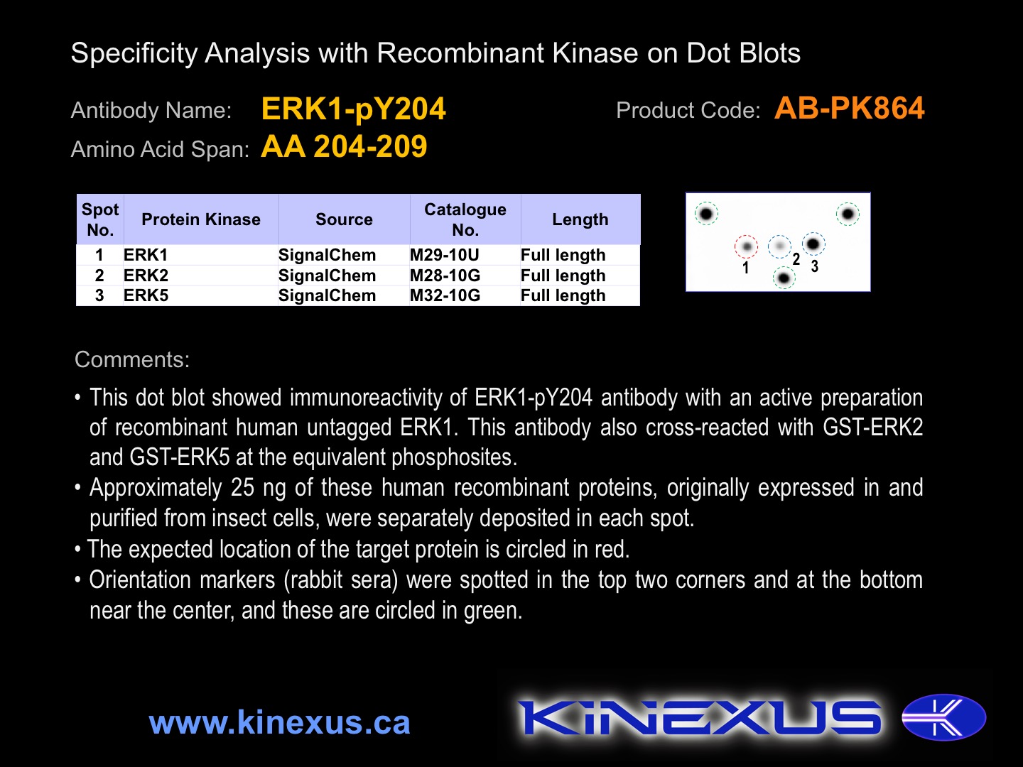
Figure 1. Dot blotting ERK1-pY204 antibody with recombinant purified proteins.
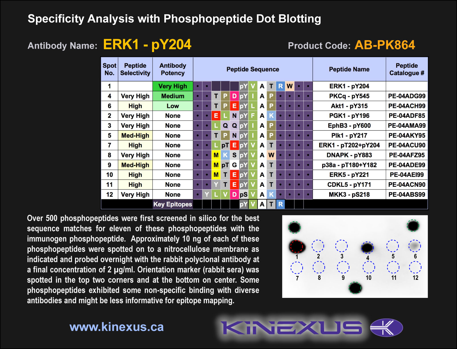
Figure 2. Epitope mapping of ERK1-pY204 antibody with similar phosphopeptides on dot blots.
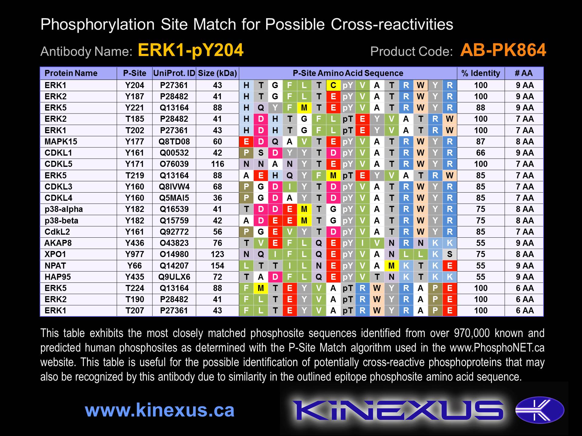
Figure 3. Identification of phosphosites related to ERK1-pY204.

© Kinexus Bioinformatics Corporation 2017