
Product Name: EGFR
Product Number: AB-NK052-3
| Size: | 25 µg | Price: | 89.00 | |
| $US |
Target Full Name: Epidermal growth factor receptor-tyrosine kinase
Target Alias: EGFR; Epidermal growth factor receptor; ErbB-1; ErbB, mENA; HER1; Receptor tyrosine-protein kinase ErbB-1; V-erb-b oncogene homologue; PIG61; CCDS5514.1; ENSG00000146648
Product Type Specific: Protein kinase pan-specific antibody
Antibody Code: NK052-3
Antibody Target Type: Pan-specific
Protein UniProt: P00533
Protein SigNET: P00533
Antibody Type: Polyclonal
Antibody Host Species: Rabbit
Antibody Immunogen Source: Human EGFR (ErbB1) sequence peptide Cat. No.:
Antibody Immunogen Sequence: SNMSMDFQNHLGSC, VIQGDERMHLPSPT, EYLRVAPQSSEFIGA
Antibody Immunogen Description: Corresponds to amino acid residues S174 to S187 + V980 to T993 + E1196 to A1210
Production Method: The immunizing peptide was produced by solid phase synthesis on a multipep peptide synthesizer and purified by reverse-phase hplc chromatography. Purity was assessed by analytical hplc and the amino acid sequence confirmed by mass spectrometry analysis. This peptide was coupled to KLH prior to immunization into rabbits. New Zealand White rabbits were subcutaneously injected with KLH-coupled immunizing peptide every 4 weeks for 4 months. The sera from these animals was applied onto an agarose column to which the immunogen peptide was thio-linked. Antibody was eluted from the column with 0.1 M glycine, pH 2.5. Subsequently, the antibody solution was neutralized to pH 7.0 with saturated Tris.
Antibody Modification: Unconjugated. Contact KInexus if you are interest in having the antibody biotinylated or coupled with fluorescent dyes.
Storage Buffer: Phosphate buffered saline pH 7.4, 0.05% Thimerasol
Storage Conditions: For long term storage, keep frozen at -40°C or lower. Stock solution can be kept at +4°C for more than 3 months. Avoid repeated freeze-thaw cycles.
Product Use: Western blotting | Antibody microarray
Antibody Dilution Recommended: 2 µg/ml for immunoblotting
Antibody Species Reactivity: Human; Mouse; Rat
Antibody Positive Control: The observed molecular mass of the processed target protein on SDS-PAGE gels is reported to be around 160-180 kDa.
Related Product 1: EGFR pan-specific antibody (Cat. No.: AB-NK052-4P)
Related Product 2: EGFR-1 pan-specific antibody (Cat. No.: AB-NK052-5)
Related Product 3: EGFR-2 pan-specific antibody (Cat. No.: AB-NK052-6)
Related Product 4: EGFR-3 pan-specific antibody (Cat. No.: AB-NK052-4)
Related Product 5: EGFR-4 pan-specific antibody (Cat. No.: AB-NK052-7)
Related Product 6: EGFR-5 pan-specific antibody (Cat. No.: AB-NK052-8)
Related Product 7: EGFR-pY1069 phosphosite-specific antibody (Cat. No.: AB-PK599)
Related Product 8: EGFR-pY1110 phosphosite-specific antibody (Cat. No.: AB-PK600)
Related Product 9: EGFR-pY1172 phosphosite-specific antibody (Cat. No.: AB-PK601)
Related Product 10: EGFRSubtide - EGFR protein kinase substrate peptide
Related Product 10: EGFRSubtide - EGFR protein kinase substrate peptide
Scientific Background: EGFR (ERBB1, HER1) is a protein-tyrosine kinase of the TK group and EGFR family. EGFR regulates cell proliferation, division, motility, survival, and has other roles in tissue development. It is activated by binding epidermal growth factor (EGF) and transforming growth factor-alpha (TGFa), which induce dimerization and autophosphorylation. Phosphorylation of Y270 stimulates interaction with EGFR. Phosphorylation of Y869 increases phosphotransferase activity and induces interaction with COX2. Phosphorylation of Y1016 increases phosphotransferase activity and induces interaction with GrbB2, PLCg1, PTPN11 (SHP2), RasGAP, and Vav2. Phosphorylation of Y1069 increases phosphotransferase activity and induces interaction with Cbl. Phosphorylation of Y1092 induces interaction with PLCg1, PTPN6 (SHP1) and Ras-GAP. Phosphorylation of Y1110 induces interaction with Ras-GAP. Phosphorylation of Y1172 increases phosphotransferase activity and induces interaction with Dok1, EGFR, RasGAP, PTPN11 (SHP2), and Vav2. Phosphorylation of Y1197 increases phosphotransferase activity and induces interaction with Cbl, EGR, PLCg1, RasGAP, SH3KBP1, Shc1, and PTPN6 (SHP1). EGFR is inhibited by phosphorylation at T678,S695 and S1026. Phosphorylation of T693, S695, S1070, S1071, and S1190 contributes to receptor internalization. EGFR is a known oncoprotein (OP). Cancer-related mutations in human tumours point to a gain of function of the protein kinase. Constitutive activation of EGFR kinase activity has been seen with the mutations of V689M, E1005R+D1006K. The active form of the protein kinase normally acts to promote tumour cell proliferation.
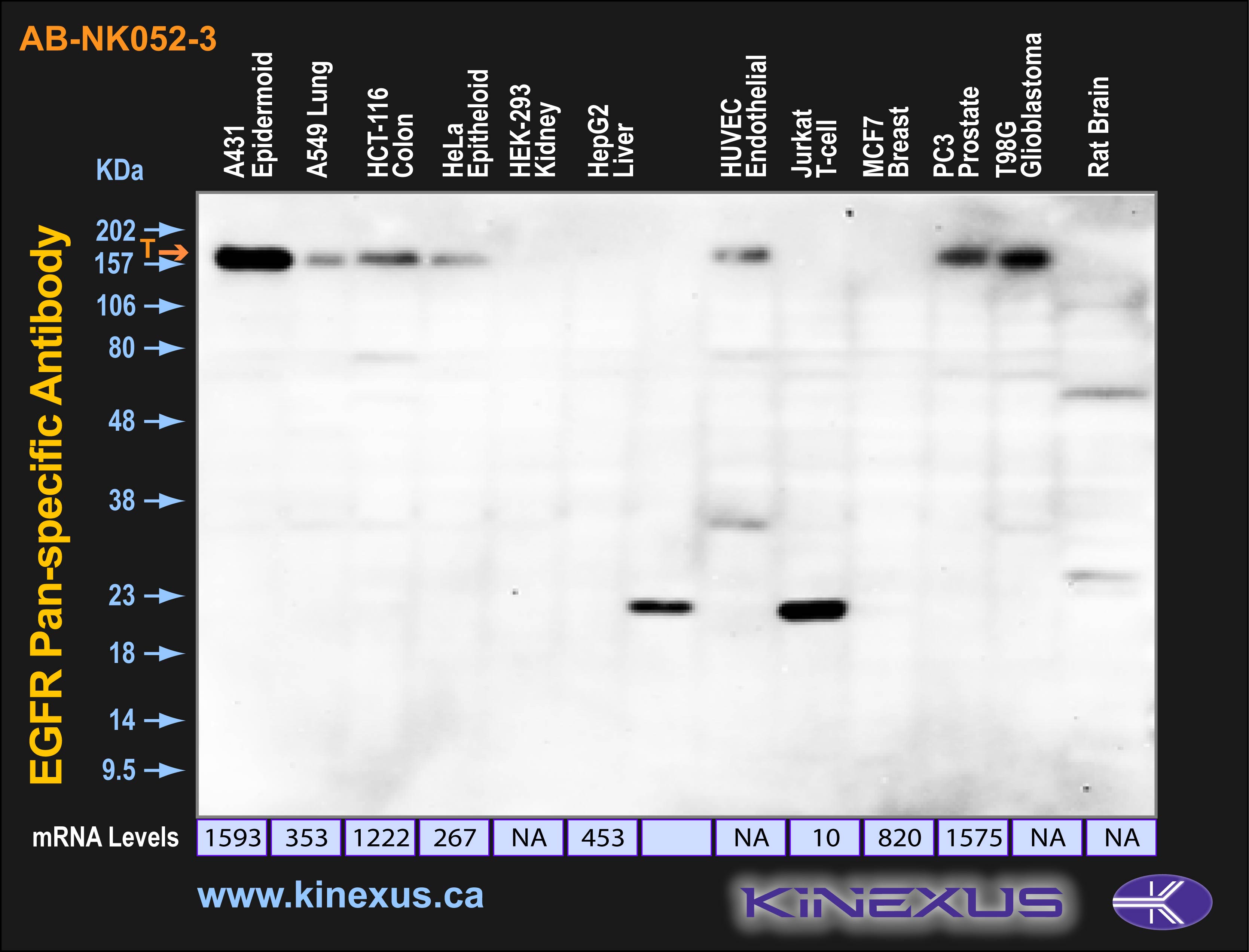
Figure 1. Immunoblotting of various cell lines with NK052-3 antibody at a 500 X dilution. The position of the full size target protein EGFR is indicated. The expected molecular mass of EGFR is 180 kDa on SDS-PAGE gels with a calculated mass of 134 kDa. Each lane was loaded with 20 µg of cell lysate protein.

© Kinexus Bioinformatics Corporation 2017