
Product Name: CDK1-pT161
Product Number: AB-PK561
| Size: | 25 µg | Price: | 89.00 | |
| $US |
Target Full Name: Cyclin-dependent protein-serine kinase 1; Cell division control protein 2 homologue
Target Alias: Cdc2; CDC28; CDC28A; CDC2A; Cell division control protein 2; Cell division cycle 2, G1 to S and G2 to M; Cyclin-dependent kinase 1; P34 protein kinase; Kinase Cdc2; MPF; DKFZp686L20222; MGC111195; ENSG00000170312
Product Type Specific: Protein kinase phosphosite-specific antibody
Antibody Code: PK561
Antibody Target Type: Phosphosite-specific
Antibody Phosphosite: T161
Protein UniProt: P06493
Protein SigNET: P06493
Antibody Type: Polyclonal
Antibody Host Species: Rabbit
Antibody Immunogen Source: Human CDK1 (CDC2) sequence peptide Cat. No.: PE-04AJG95
Antibody Immunogen Sequence: RVY(pT)HEV(bA)C
Antibody Immunogen Description: Corresponds to amino acid residues R158 to V164; In the protein kinase catalytic domain activation T loop region between subdomains VII and VIII.
Production Method: The immunizing peptide was produced by solid phase synthesis on a multipep peptide synthesizer and purified by reverse-phase hplc chromatography. Purity was assessed by analytical hplc and the amino acid sequence confirmed by mass spectrometry analysis. This peptide was coupled to KLH prior to immunization into rabbits. New Zealand White rabbits were subcutaneously injected with KLH-coupled immunizing peptide every 4 weeks for 4 months. The sera from these animals was applied onto an agarose column to which the immunogen peptide was thio-linked. Antibody was eluted from the column with 0.1 M glycine, pH 2.5. Subsequently, the antibody solution was neutralized to pH 7.0 with saturated Tris.This antibody was also subject to negative purification over phosphotyrosine-agarose.
Antibody Modification: Unconjugated. Contact KInexus if you are interest in having the antibody biotinylated or coupled with fluorescent dyes.
Antibody Modification: Unconjugated. Contact KInexus if you are interest in having the antibody biotinylated or coupled with fluorescent dyes.
Antibody Concentration: 1 mg/ml
Storage Buffer: Phosphate buffered saline pH 7.4, 0.05% Thimerasol
Storage Conditions: For long term storage, keep frozen at -40°C or lower. Stock solution can be kept at +4°C for more than 3 months. Avoid repeated freeze-thaw cycles.
Product Use: Western blotting | Antibody microarray
Antibody Dilution Recommended: 2 µg/ml for immunoblotting
Antibody Potency: Weak immunoreactivity of a target-sized protein by Western blotting in maturing sea star oocytes. Very strong immunoreactivity with immunogen peptide on dot blots.
Antibody Species Reactivity: Human
Antibody Positive Control: The observed molecular mass of the processed target protein on SDS-PAGE gels is reported to be around 30-35 kDa.
Antibody Specificity: Very high
Antibody Cross Reactivity: No cross-reactive proteins detected in HeLa and A431 cells. A medium ~37 kDa cross-reactive protein was detected in sea star oocytes.
Related Product 1: CDK1-pT161 blocking peptide
Related Product 2: CDC2-CT (CDK1-1) pan-specific antibody (Cat. No.: AB-NK025-4)
Related Product 3: CDK1-X pan-specific antibody (Cat. No.: AB-NK025-7)
Related Product 4: CDK1-pT14 phosphosite-specific antibody (Cat. No.: AB-PK559)
Related Product 5: CDK1-pT14+pY15 phosphosite-specific antibody (Cat. No.: AB-PK560)
Related Product 6: CDK1-pY15 phosphosite-specific antibody (Cat. No.: AB-PK562)
Related Product 7: CDK1-pY19 phosphosite-specific antibody (Cat. No.: AB-PK563)
Related Product 8: CDK1-3 Selectide - CDK1 (CDC2) protein kinase substrate peptide
Related Product 6: CDK1-pY15 phosphosite-specific antibody (Cat. No.: AB-PK562)
Related Product 7: CDK1-pY19 phosphosite-specific antibody (Cat. No.: AB-PK563)
Related Product 8: CDK1-3 Selectide - CDK1 (CDC2) protein kinase substrate peptide
Scientific Background: CDK1 (CDC2) is a protein-serine/threonine kinase of the CMGC group and CDK family. It plays an essential role in cell cycle control in eukaryotic cells by regulating the centrosome cycle, mitotic onset, G2-M phase transition, G1 progression, and G1-S phase transition through an association with various interphase cyclin proteins. Phosphorylation events at T14 or Y15 on the protein are inactivating, while phosphorylation at T161 is stimulatory. CDK1 appears to be a tumour requiring protein (TRP). Gain-of-function mutations in the CDK1 gene have been linked to several forms of cancer, indicating an oncogenic role for the CDK1 protein. Cells transformed with the oncogene MYC undergo apoptosis when treated with small-molecule CDK1 inhibitors. Elevated expression of CDK1 has been reported as a diagnostic marker for cancer progression in esophageal adenocarcinoma, potentially reflecting the role of the CDK1 protein in tumourigenesis. CDK1 expression can be used as a prognostic indicator for early breast cancer. For example, breast cancer tumours with high expression of CDK1 are correlated with a significantly lower 5-year patient survival rate (66. 9%) than tumours that have low levels of CDK1 expression (84. 2%).
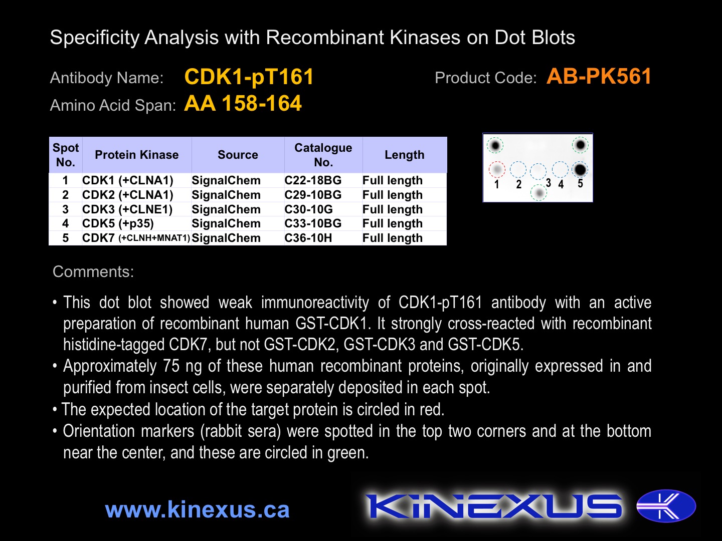
Figure 1. Dot blotting CDK1-pT161 antibody with recombinant purified proteins.
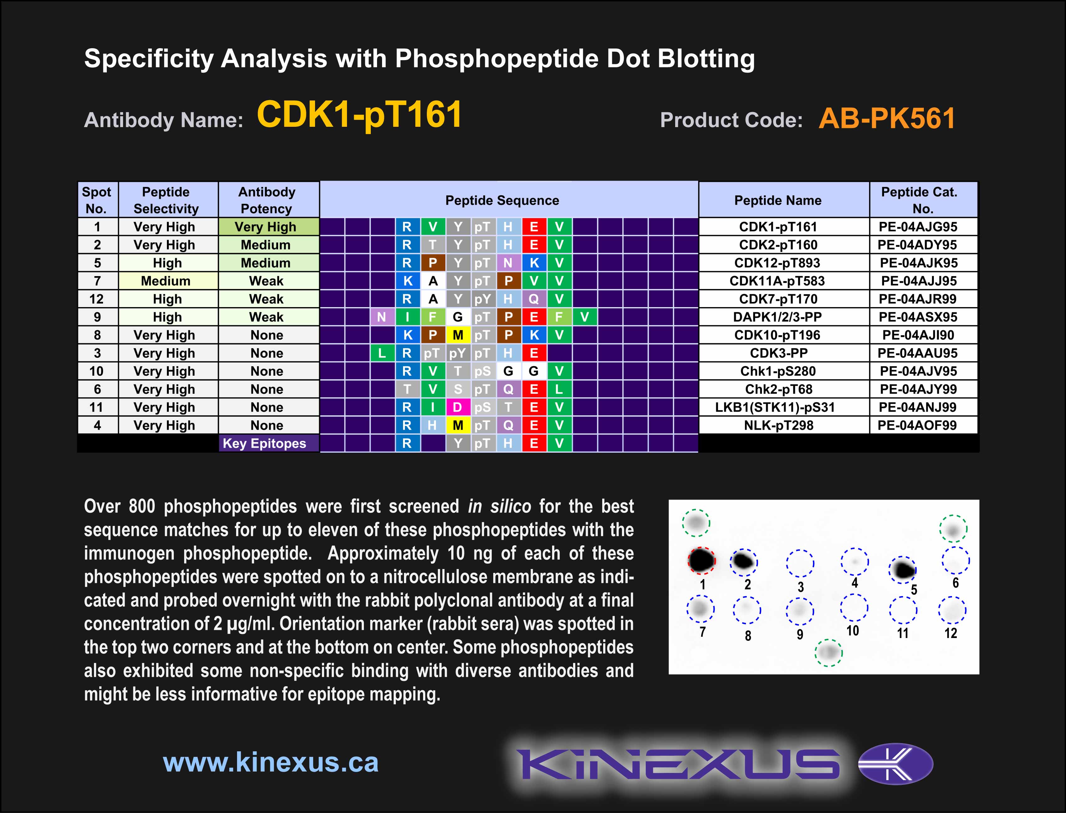
Figure 2. Epitope mapping of CDK1-pT161 antibody with similar phosphopeptides on dot blots.
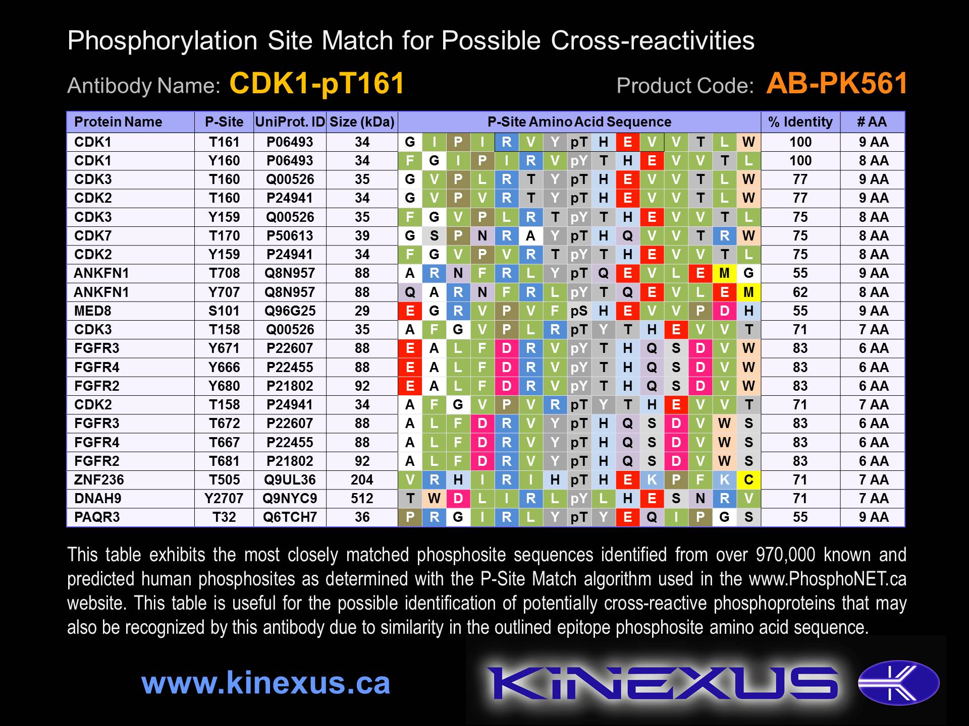
Figure 3. Identification of phosphosites related to CDK1-pT161.

© Kinexus Bioinformatics Corporation 2017