
Product Name: EphA2-pY588
Product Number: AB-PK606
| Size: | 25 µg | Price: | 89.00 | |
| $US |
Target Full Name: Ephrin type-A receptor 2 protein-tyrosine kinase
Target Alias: ECK; Eph receptor A2; Epithelial cell kinase; Kinase EphA2; MPK-5; SEK2; SEK-2; Tyrosine-protein kinase receptor ECK; RCC2; CCDS169.1; ENSG00000142627
Product Type Specific: Protein kinase phosphosite-specific antibody
Antibody Code: PK606
Antibody Target Type: Phosphosite-specific
Antibody Phosphosite: Y588
Protein UniProt: P29317
Protein SigNET: P29317
Antibody Type: Polyclonal
Antibody Host Species: Rabbit
Antibody Immunogen Source: Human EphA2 sequence peptide Cat. No.: PE-04ALW99
Antibody Immunogen Sequence: LKT(pY)VDP(bA)C
Antibody Immunogen Description: Corresponds to amino acid residues L585 to P591; In the EphA2-TM domain located prior to the kinase catalytic domain. One of the major in vivo sites of phosphorylation in EphA2.
Antibody Immunogen Sequence: LKT(pY)VDP(bA)C
Antibody Immunogen Description: Corresponds to amino acid residues L585 to P591; In the EphA2-TM domain located prior to the kinase catalytic domain. One of the major in vivo sites of phosphorylation in EphA2.
Production Method: The immunizing peptide was produced by solid phase synthesis on a multipep peptide synthesizer and purified by reverse-phase hplc chromatography. Purity was assessed by analytical hplc and the amino acid sequence confirmed by mass spectrometry analysis. This peptide was coupled to KLH prior to immunization into rabbits. New Zealand White rabbits were subcutaneously injected with KLH-coupled immunizing peptide every 4 weeks for 4 months. The sera from these animals was applied onto an agarose column to which the immunogen peptide was thio-linked. Antibody was eluted from the column with 0.1 M glycine, pH 2.5. Subsequently, the antibody solution was neutralized to pH 7.0 with saturated Tris.This antibody was also subject to negative purification over phosphotyrosine-agarose.
Antibody Modification: Unconjugated. Contact KInexus if you are interest in having the antibody biotinylated or coupled with fluorescent dyes.
Antibody Modification: Unconjugated. Contact KInexus if you are interest in having the antibody biotinylated or coupled with fluorescent dyes.
Antibody Concentration: 1 mg/ml
Storage Buffer: Phosphate buffered saline pH 7.4, 0.05% Thimerasol
Storage Conditions: For long term storage, keep frozen at -40°C or lower. Stock solution can be kept at +4°C for more than 3 months. Avoid repeated freeze-thaw cycles.
Product Use: Western blotting | Antibody microarray
Antibody Dilution Recommended: 2 µg/ml for immunoblotting
Antibody Potency: Very strong immunoreactivity with immunogen peptide on dot blots.
Antibody Species Reactivity: Human
Antibody Positive Control: The observed molecular mass of the processed target protein on SDS-PAGE gels is reported to be around 115-130 kDa.
Antibody Specificity: Very high
Antibody Cross Reactivity: No significant cross-reactive proteins detected in HeLa cells, except for a weak ~75 kDa cross-reactive protein.
Related Product 1: EphA2-pY588 blocking peptide
Related Product 2: EphA1-pY781 phosphosite-specific antibody (Cat. No.: AB-PK605)
Related Product 3: EphA3-pY779 phosphosite-specific antibody (Cat. No.: AB-PK608)
Scientific Background: EphA2 is a protein-tyrosine kinase of the TK group and Eph family. It is a receptor that displays promiscuous binding to membrane-bound ligands of the ephrin-A family on adjacent cells. It is activated by binding ephrin-A1, A2, A3, A4 or A5. Phosphorylation of Y735 increases phosphotranserase activity and interaction with PIK3R1. EphA2 activation regulates integrin-dependent adhesion, cellular migration, proliferation, and differentiation. EphA2 also participates in bone remodeling by directly regulating both osteoclastogenesis and osteoblastogenesis. EphA2 may be an oncoprotein (OP). Significantly elevated EphA2 expression has been observed in many types of human cancer and appears to be involved in mediating communication between RAS-PI3K/Akt and RAS-MAPK intracellular signalling pathways, which are both known to promote tumourigenesis. Additionally, EphA2 is required for UV-radiation induced apoptosis. UV-radiation is a potent carcinogen responsible for the development of melanoma. However, further characterization of melanoma cell lines revealed either a pro-apoptotic or anti-apoptotic role for EphA2 dependent upon the cellular context. Additionally, studies performed in breast cancer cell lines revealed a link between EphA2 expression levels and changes in cytoskeletal morphology and the recruitment of disintegrin and metalloprotease-10 (MMP10) enzymes, thus suggesting a role for theEphA2 protein in the promotion of tumour invasion and metastasis.
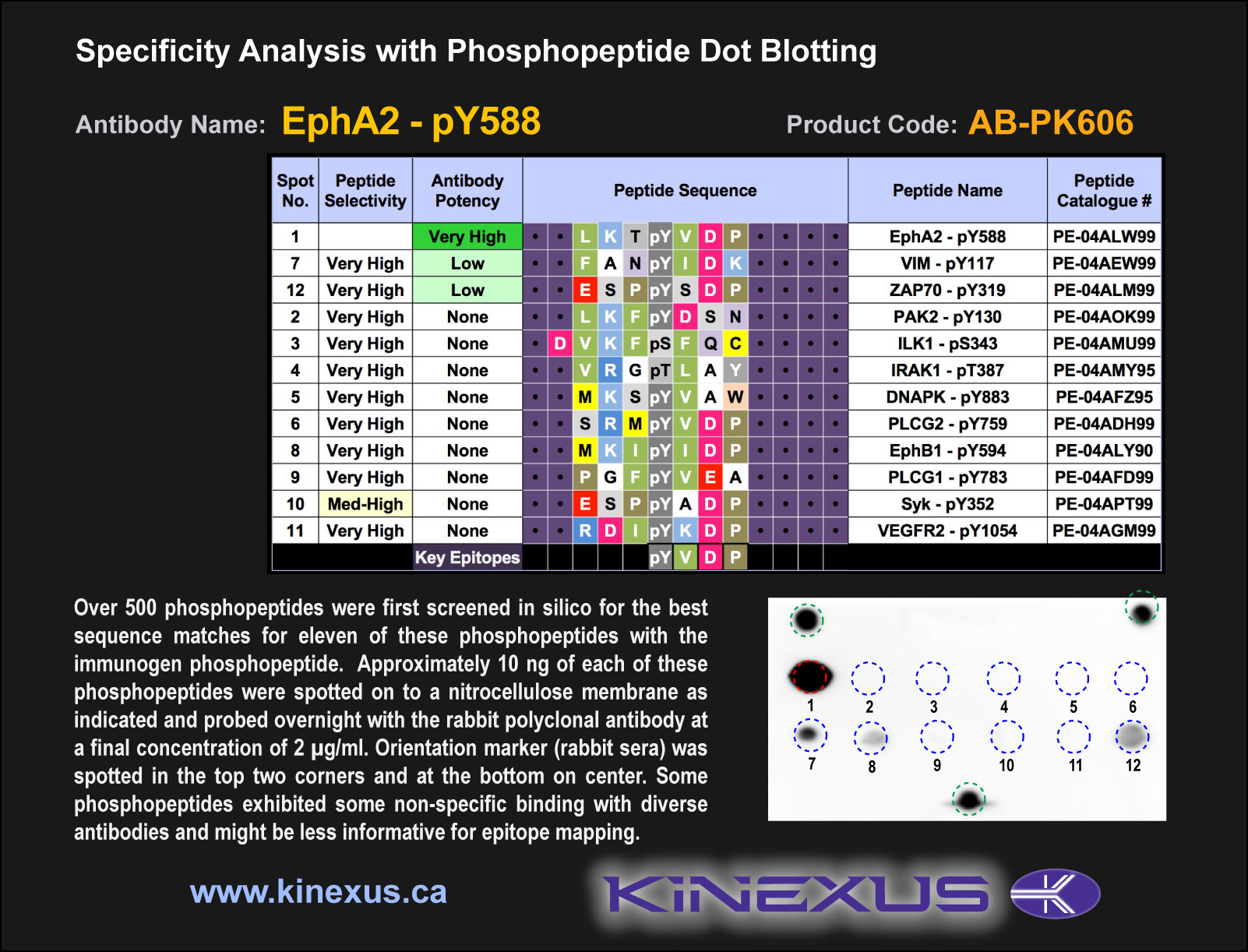
Figure 1. Epitope mapping of EphA2-pY588 antibody with similar phosphopeptides on dot blots.
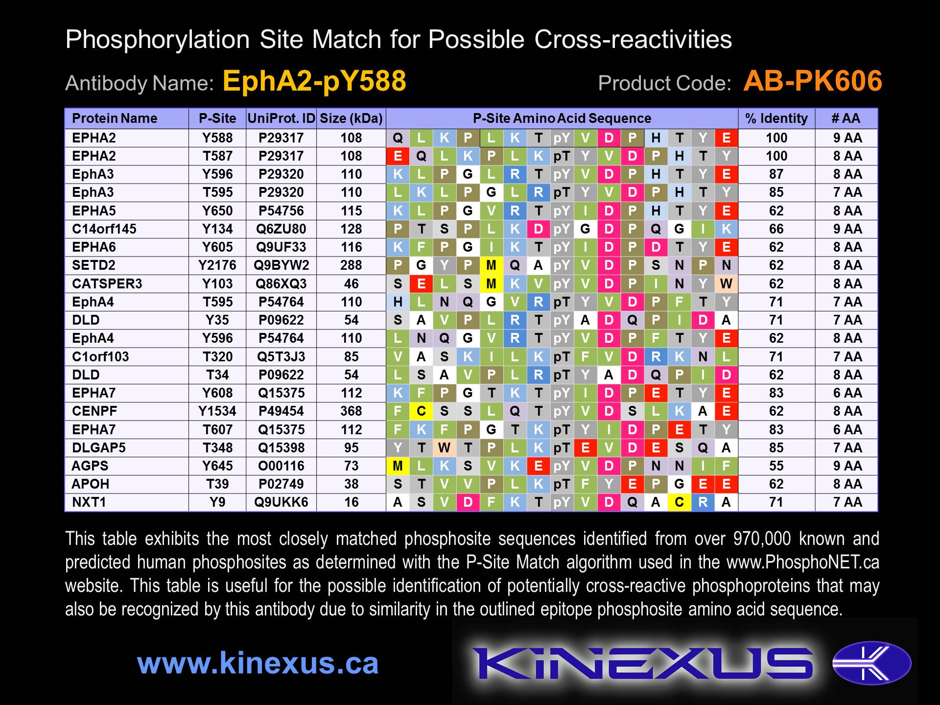
Figure 2. Identification of phosphosites related to EphA2-pY588.

© Kinexus Bioinformatics Corporation 2017