
Product Name: MELK-pY438
Product Number: AB-PK701
| Size: | 25 µg | Price: | 89.00 | |
| $US |
Target Full Name: Maternal embryonic leucine zipper kinase
Target Alias: hMELK; hPK38; KIAA0175; Maternal embryonic leucine zipper kinase; MELK; pEg3 kinase; Protein kinase PK38
Product Type Specific: Protein kinase phosphosite-specific antibody
Antibody Code: PK701
Antibody Target Type: Phosphosite-specific
Antibody Phosphosite: Y438
Protein UniProt: Q14680
Protein SigNET: Q14680
Antibody Type: Polyclonal
Antibody Host Species: Rabbit
Antibody Immunogen Source: Human MELK sequence peptide Cat. No.: PE-04AOA01
Antibody Immunogen Sequence: NEE(pY)FMF(bA)C
Antibody Immunogen Description: Corresponds to amino acid residues N435 to F441; In the C-terminal third of the protein kinase. This is the major in vivo phosphorylation site in MELK.
Antibody Immunogen Description: Corresponds to amino acid residues N435 to F441; In the C-terminal third of the protein kinase. This is the major in vivo phosphorylation site in MELK.
Production Method: The immunizing peptide was produced by solid phase synthesis on a multipep peptide synthesizer and purified by reverse-phase hplc chromatography. Purity was assessed by analytical hplc and the amino acid sequence confirmed by mass spectrometry analysis. This peptide was coupled to KLH prior to immunization into rabbits. New Zealand White rabbits were subcutaneously injected with KLH-coupled immunizing peptide every 4 weeks for 4 months. The sera from these animals was applied onto an agarose column to which the immunogen peptide was thio-linked. Antibody was eluted from the column with 0.1 M glycine, pH 2.5. Subsequently, the antibody solution was neutralized to pH 7.0 with saturated Tris.This antibody was also subject to negative purification over phosphotyrosine-agarose.
Antibody Modification: Unconjugated. Contact KInexus if you are interest in having the antibody biotinylated or coupled with fluorescent dyes.
Antibody Modification: Unconjugated. Contact KInexus if you are interest in having the antibody biotinylated or coupled with fluorescent dyes.
Antibody Concentration: 0.5 mg/ml
Storage Buffer: Phosphate buffered saline pH 7.4, 0.05% Thimerasol
Storage Conditions: For long term storage, keep frozen at -40°C or lower. Stock solution can be kept at +4°C for more than 3 months. Avoid repeated freeze-thaw cycles.
Product Use: Western blotting | Antibody microarray
Antibody Dilution Recommended: 2 µg/ml for immunoblotting
Antibody Potency: Medium immunoreactivity with recombinant human MELK on protein dot blots.
Antibody Species Reactivity: Human
Antibody Positive Control: The observed molecular mass of the processed target protein on SDS-PAGE gels is reported to be around 70-75 kDa.
Antibody Specificity: Very high
Antibody Cross Reactivity: No significant cross-reactive proteins detected in phenylarsine oxide (PAO)+vanadate-treated HeLa cells, EGF-treated A431 cells and insulin-treated MCF7 cells, when these cells were homogenized in SDS-PAGE sample buffer.
Related Product 1: MELK-pY438 blocking peptide
Related Product 2: MELK pan-specific antibody (Cat. No.: AB-NK229-1)
Related Product 3: MELK pan-specific antibody (Cat. No.: AB-NK229-2P)
Related Product 4: MELK-2 pan-specific antibody (Cat. No.: AB-NK229-3)
Related Product 5: MELK-pY438 phosphosite-specific antibody (Cat. No.: AB-PK701)
Scientific Background: MELK is a protein-serine/threonine kinase of the CAMK group and CAMKL family. It can occur in 8 human isoforms ranging from 74.642 to 52.528 kDa in size. It is involved in various processes such as cell cycle regulation, self-renewal of stem cells, apoptosis and splicing regulation. It is activated by autophosphorylation of the T-loop at T167 and S171: in contrast to other members of the SNF1 subfamily, phosphorylation at T167 is not mediated by STK11/LKB1 but via autophosphorylation instead. Phosphorylation at T478 induces interaction with NIPP-1. It exhibits a broad substrate specificity with targets that include BCL2L14, CDC25B, MAP3K5/ASK1 and ZNF622. It is an activator of apoptosis by phosphorylating and activating MAP3K5/ASK1, and its phosphorylation of CDC25B, promoting localization of CDC25B to the centrosome and the spindle poles during mitosis. It is required for proliferation of embryonic and postnatal multipotent neural progenitors. It phosphorylates and inhibits BCL2L14, possibly leading to affect mammary carcinogenesis by mediating inhibition of its pro-apoptotic function. It inhibits spliceosome assembly during mitosis by phosphorylating ZNF622, which contributes to its redirection to the nucleus. It interacts with ZNF622 and PPP1R8. It is expressed in placenta, kidney, thymus, testis, ovary and intestine, and is up-regulated in many cancers cells.
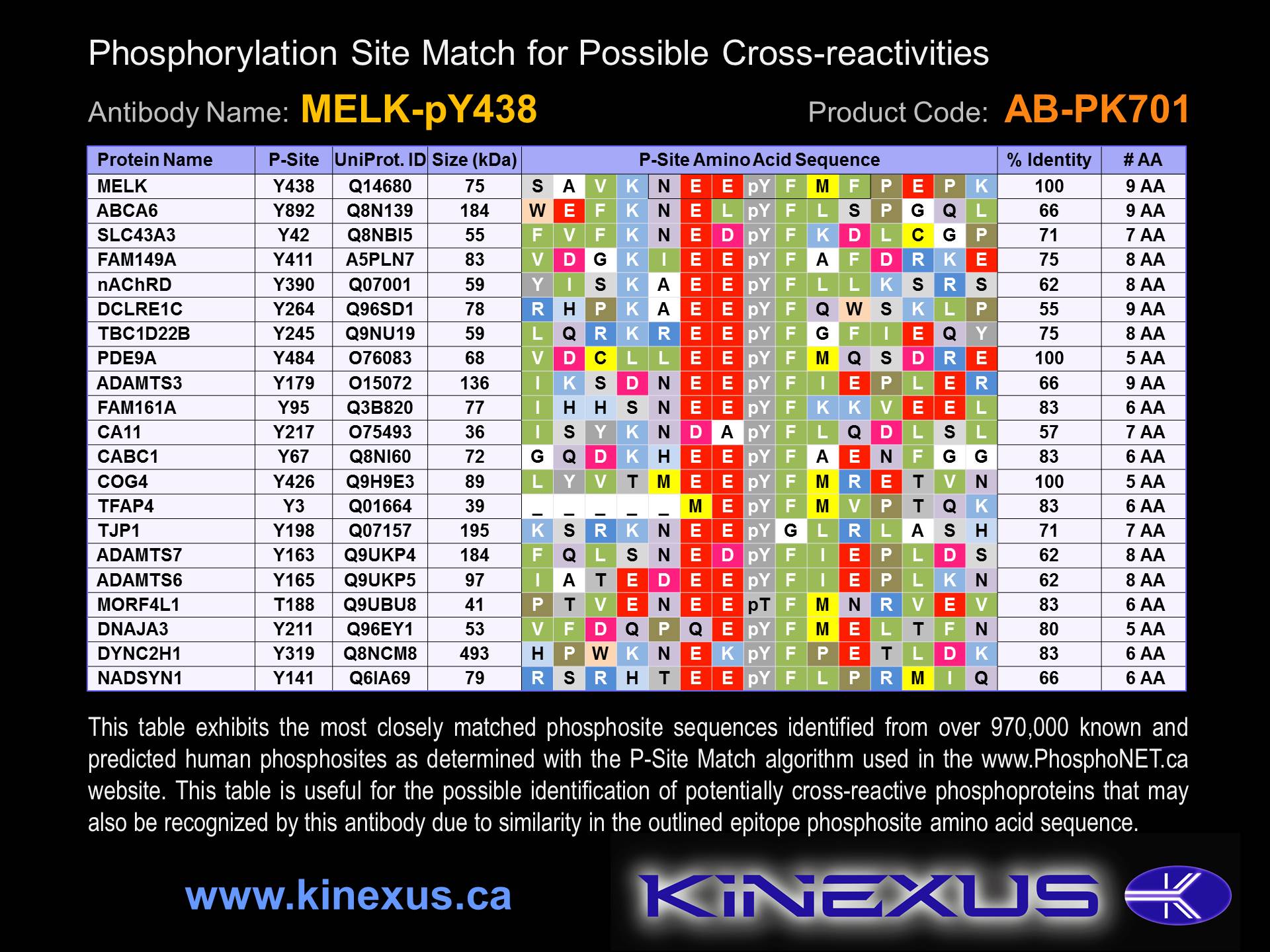
Figure 1. Identification of phosphosites related to MELK-pY438.

© Kinexus Bioinformatics Corporation 2017