
Product Name: TAK1-pT184+pT187
Product Number: AB-PK825
| Size: | 25 µg | Price: | 89.00 | |
| $US |
Target Full Name: TGF-beta-activated protein-serine kinase 1; Mitogen-activated protein kinase kinase kinase 7
Target Alias: M3K7; MAP3K7; MEKK7; TGF1a; TGF-beta-activated kinase 1; TGF1a; ENSG00000135341
Product Type Specific: Protein kinase phosphosite-specific antibody
Antibody Code: PK825
Antibody Target Type: Phosphosite-specific
Antibody Phosphosite: T184+T187
Protein UniProt: O43318
Protein SigNET: O43318
Antibody Type: Polyclonal
Antibody Host Species: Rabbit
Antibody Immunogen Source: Human TAK1 sequence peptide Cat. No.: PE-04APW99
Antibody Immunogen Sequence: DIQ(pT)HM(pT)NNK(bA)C
Antibody Immunogen Description: Corresponds to amino acid residues D181 to K190; In protein kinase catalytic domain activation T-loop between subdomains VII and VIII.
Antibody Immunogen Sequence: DIQ(pT)HM(pT)NNK(bA)C
Antibody Immunogen Description: Corresponds to amino acid residues D181 to K190; In protein kinase catalytic domain activation T-loop between subdomains VII and VIII.
Production Method: The immunizing peptide was produced by solid phase synthesis on a multipep peptide synthesizer and purified by reverse-phase hplc chromatography. Purity was assessed by analytical hplc and the amino acid sequence confirmed by mass spectrometry analysis. This peptide was coupled to KLH prior to immunization into rabbits. New Zealand White rabbits were subcutaneously injected with KLH-coupled immunizing peptide every 4 weeks for 4 months. The sera from these animals was applied onto an agarose column to which the immunogen peptide was thio-linked. Antibody was eluted from the column with 0.1 M glycine, pH 2.5. Subsequently, the antibody solution was neutralized to pH 7.0 with saturated Tris.This antibody was also subject to negative purification over phosphotyrosine-agarose.
Antibody Modification: Unconjugated. Contact KInexus if you are interest in having the antibody biotinylated or coupled with fluorescent dyes.
Antibody Modification: Unconjugated. Contact KInexus if you are interest in having the antibody biotinylated or coupled with fluorescent dyes.
Antibody Concentration: 0.5 mg/ml
Storage Buffer: Phosphate buffered saline pH 7.4, 0.05% Thimerasol
Storage Conditions: For long term storage, keep frozen at -40°C or lower. Stock solution can be kept at +4°C for more than 3 months. Avoid repeated freeze-thaw cycles.
Product Use: Western blotting | Antibody microarray
Antibody Dilution Recommended: 2 µg/ml for immunoblotting
Antibody Potency: Medium immunoreactivity of a target-sized protein by Western blotting in Jurkat and MCF7 cells. Medium immunoreactivity with immunogen peptide on dot blots.
Antibody Species Reactivity: Human
Antibody Positive Control: The observed molecular mass of the processed target protein on SDS-PAGE gels is reported to be around 67-85 kDa.
Antibody Specificity: Medium
Antibody Cross Reactivity: In HepG2 cells, very high background and antibody detects many cross-reactive proteins. In HeLa, Jurkat, and MCF7 cells, this antibody strong cross-reacts with a 40 kDa protein. In MCF7 cells, a 110-140 kDa doublet is also detected in MCF7 cells.
Related Product 1: TAK1-pT184+pT187 blocking peptide
Related Product 2: TAK1-CT (TAK1-4) pan-specific antibody (Cat. No.: AB-NK175-3)
Related Product 3: TAK1-pS439 phosphosite-specific antibody (Cat. No.: AB-PK824)
Scientific Background: TAK1 (MAP3K7, MEKK7) is a protein-serine/threonine kinase of the TKL group and MLK family. TAK1 is activated by proinflammatory cytokines and in response to physical/chemical stress, including UVR, osmotic- and oxidative stress. It is a mediator of TRAF6 and TGF-beta signal transduction, and activates IKBKB and MAPK8 in response to TRAF6 signalling. It also stimulates NFκB activation and activation of the p38 MAPK (MAPK8) pathway. It is responsible for controlling a variety of cell functions such as transcription and apoptosis. It regulates hepatic cell survival and carcinogenesis. TAK1 is important for TGF-beta1 regulation of MMP9 and the metastatic potential of certain breast cancer cell lines. It is activated by phosphorylation at T178, T184, T187 and S192. It is also activated by K63-linked polyubiquitination. TAK1 plays a role in tumour initiation, progression, and metastasis such as in breast cancer as a tumour inducer or tumour suppression depending on the cancer.
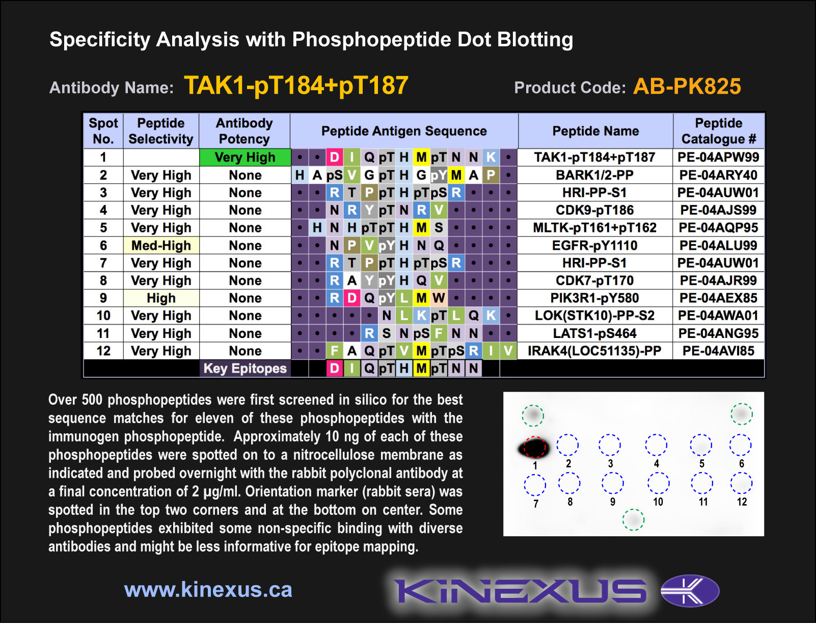
Figure 1. Epitope mapping of TAK1-pT184+pT187 antibody with similar phosphopeptides on dot blots.
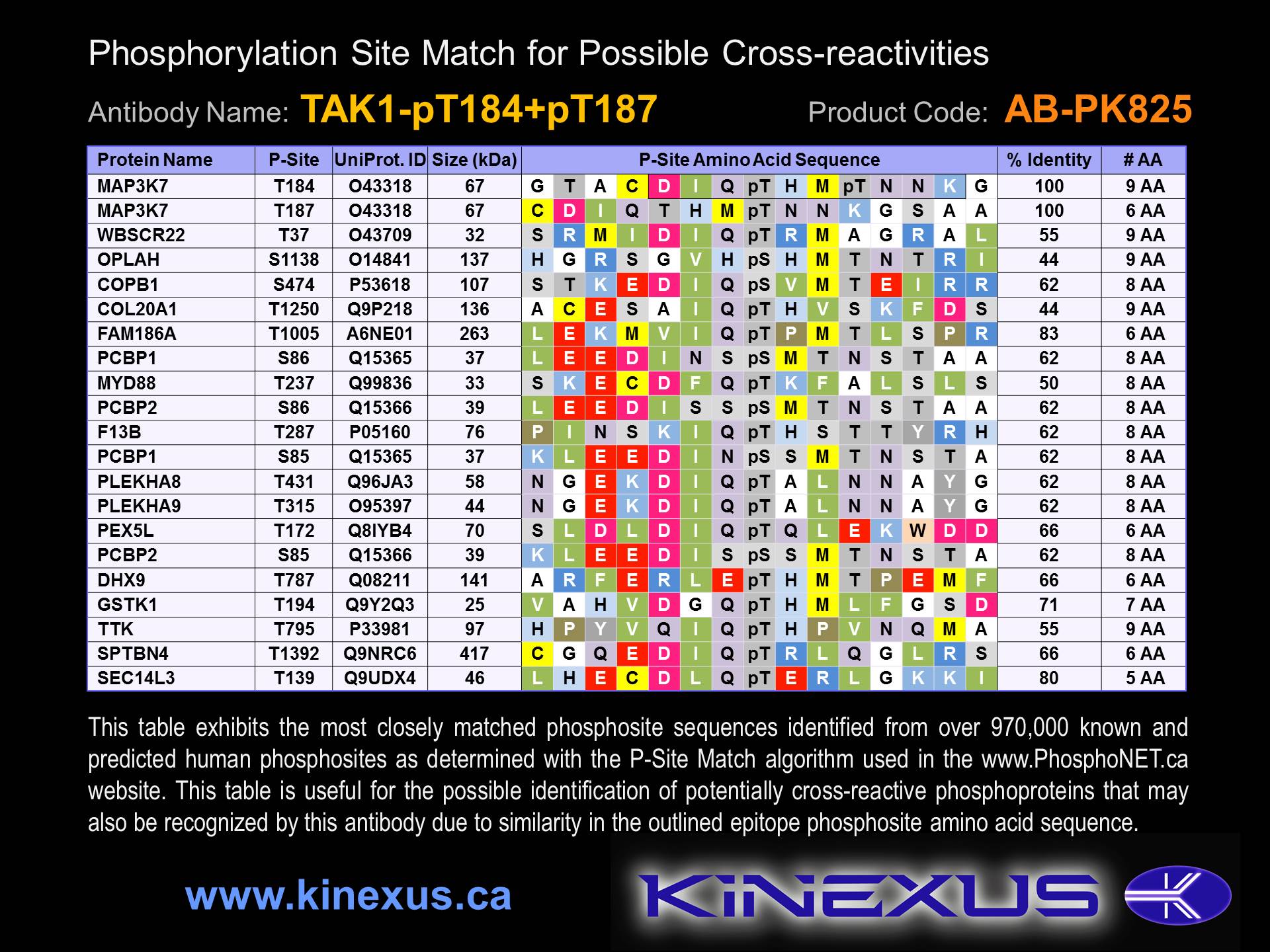
Figure 2. Identification of phosphosites related to TAK1-pT184+pT187.

© Kinexus Bioinformatics Corporation 2017