
Product Name: ErbB3-pY1328
Product Number: AB-PK618
| Size: | 25 µg | Price: | 89.00 | |
| $US |
Target Full Name: ErbB3 (HER3) receptor-tyrosine kinase
Target Alias: C-erbB3; ERB3; HER3; Kinase ErbB3; LCCS2; Receptor protein-tyrosine kinase erbB-3 precursor; Tyrosine kinase-type cell surface receptor HER3; V-erb-b2 erythroblastic leukemia viral oncogene 3; ErbB-3; c-erbB3; erbB3-S; MDA-BF-1; MGC88033; c-erbB-3; p180-ErbB3; p45-sErbB3; p85-sErbB3; ENSG00000065361; O75810; O75811; O75812; O75813; Q9BUD7; Q9NNX2; Q9NNX3
Product Type Specific: Protein kinase phosphosite-specific antibody
Antibody Code: PK618
Antibody Target Type: Phosphosite-specific
Antibody Phosphosite: Y1328
Protein UniProt: P21860
Protein SigNET: P21860
Antibody Type: Polyclonal
Antibody Host Species: Rabbit
Antibody Immunogen Source: Human ErbB3 (HER3) sequence peptide Cat. No.: PE-04AGY99
Antibody Immunogen Sequence: NPD(pY)WHS(bA)C
Antibody Immunogen Description: Corresponds to amino acid residues N1325 to S1331; In the cytoplasmic region of ErbB3 near the C-terminus after the kinase catalytic domain. This is one of the major in vivo phosphorylation sites in ErbB3.
Production Method: The immunizing peptide was produced by solid phase synthesis on a multipep peptide synthesizer and purified by reverse-phase hplc chromatography. Purity was assessed by analytical hplc and the amino acid sequence confirmed by mass spectrometry analysis. This peptide was coupled to KLH prior to immunization into rabbits. New Zealand White rabbits were subcutaneously injected with KLH-coupled immunizing peptide every 4 weeks for 4 months. The sera from these animals was applied onto an agarose column to which the immunogen peptide was thio-linked. Antibody was eluted from the column with 0.1 M glycine, pH 2.5. Subsequently, the antibody solution was neutralized to pH 7.0 with saturated Tris.This antibody was also subject to negative purification over phosphotyrosine-agarose.
Antibody Modification: Unconjugated. Contact KInexus if you are interest in having the antibody biotinylated or coupled with fluorescent dyes.
Antibody Modification: Unconjugated. Contact KInexus if you are interest in having the antibody biotinylated or coupled with fluorescent dyes.
Antibody Concentration: 1 mg/ml
Storage Buffer: Phosphate buffered saline pH 7.4, 0.05% Thimerasol
Storage Conditions: For long term storage, keep frozen at -40°C or lower. Stock solution can be kept at +4°C for more than 3 months. Avoid repeated freeze-thaw cycles.
Product Use: Western blotting | Antibody microarray
Antibody Dilution Recommended: 2 µg/ml for immunoblotting
Antibody Potency: Medium immunoreactivity of a target-sized protein by Western blotting in A431 cells and weak detection of target in HeLa cells.
Antibody Species Reactivity: Human
Antibody Positive Control: The observed molecular mass of the processed target protein on SDS-PAGE gels is reported to be around 160-200 kDa.
Antibody Specificity: High-very high
Antibody Cross Reactivity: No significant cross-reactive proteins detected in A431 and HeLa cells, except for a ~33 kDa cross-reactive protein in A431 cells.
Related Product 1: ErbB3-pY1328 blocking peptide
Related Product 2: ErbB3 pan-specific antibody (Cat. No.: AB-NK231-2P)
Related Product 3: ErbB3-1 pan-specific antibody (Cat. No.: AB-NK231-2)
Related Product 4: ErbB3-2 pan-specific antibody (Cat. No.: AB-NK231-3)
Related Product 5: ErbB3-3 pan-specific antibody (Cat. No.: AB-NK231-4)
Related Product 6: ErbB3-pY1289 phosphosite-specific antibody (Cat. No.: AB-PK616)
Related Product 7: ErbB3-pY1307 phosphosite-specific antibody (Cat. No.: AB-PK617)
Related Product 7: ErbB3-pY1307 phosphosite-specific antibody (Cat. No.: AB-PK617)
Scientific Background: ErbB3 (HER3) is a protein-tyrosine kinase of the TK group and EGFR family. It is a receptor kinase that bind and be activated by NTAK and neuregulins. It has been proposed to be deficient in phosphotransferase activity. While the kinase domain appararently lacks activity but heterodimerizes with other EGFRs permits transduction of growth signals. Phosphorylation of Y1222 and Y1260 induces interaction with PIK3C and PIK3R1. ErbB3 appears to be up-regulated in many cancer, including medulloblastomas, verrucous carcinomas, childhood medulloblastomas, carotid body tumours (CBT), and gestational trophoblastic tumours (GTD).
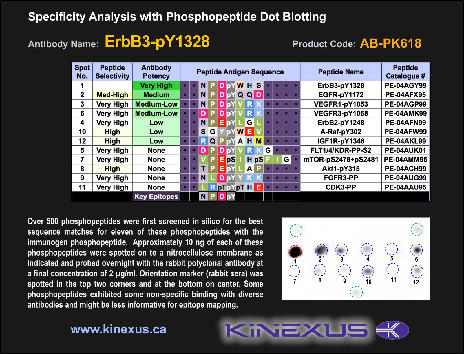
Figure 1. Epitope mapping of ErbB3-pY1328 antibody with similar phosphopeptides on dot blots.
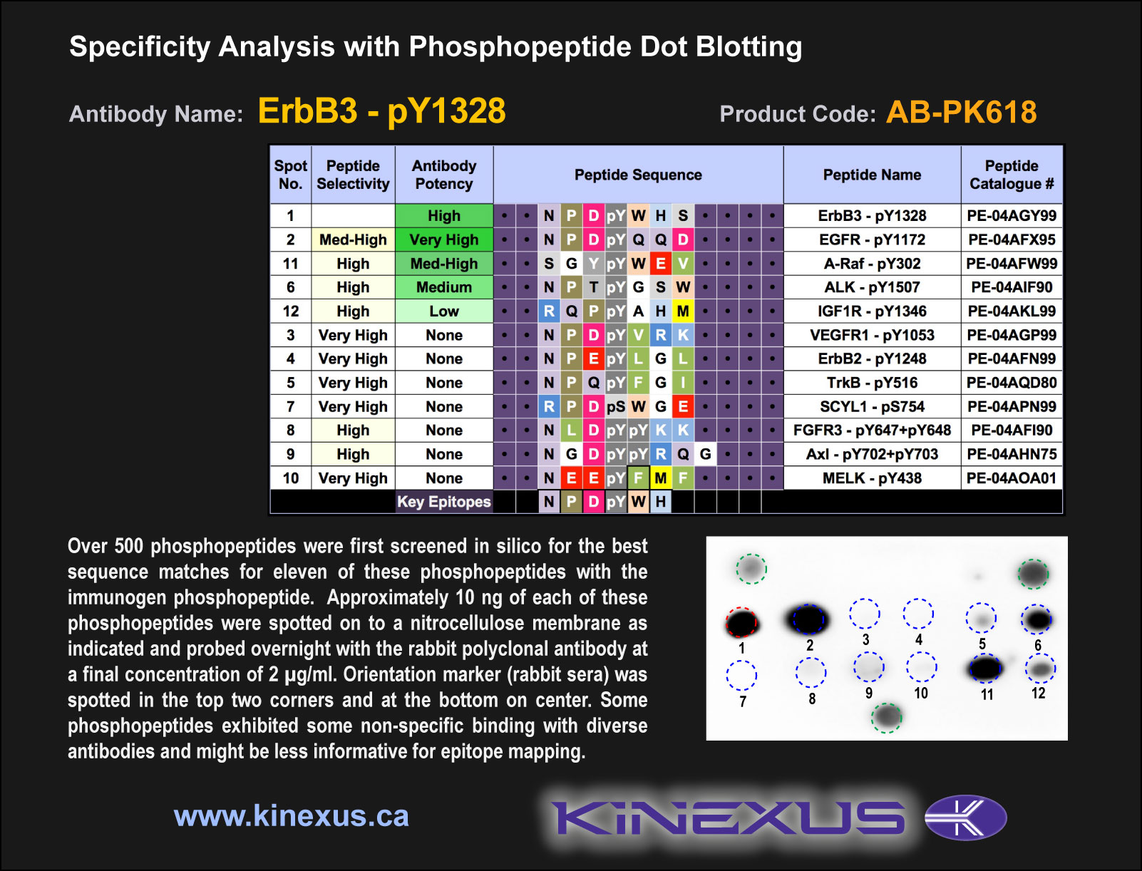
Figure 2. Epitope mapping of ErbB3-pY1328 antibody with similar phosphopeptides on dot blots.
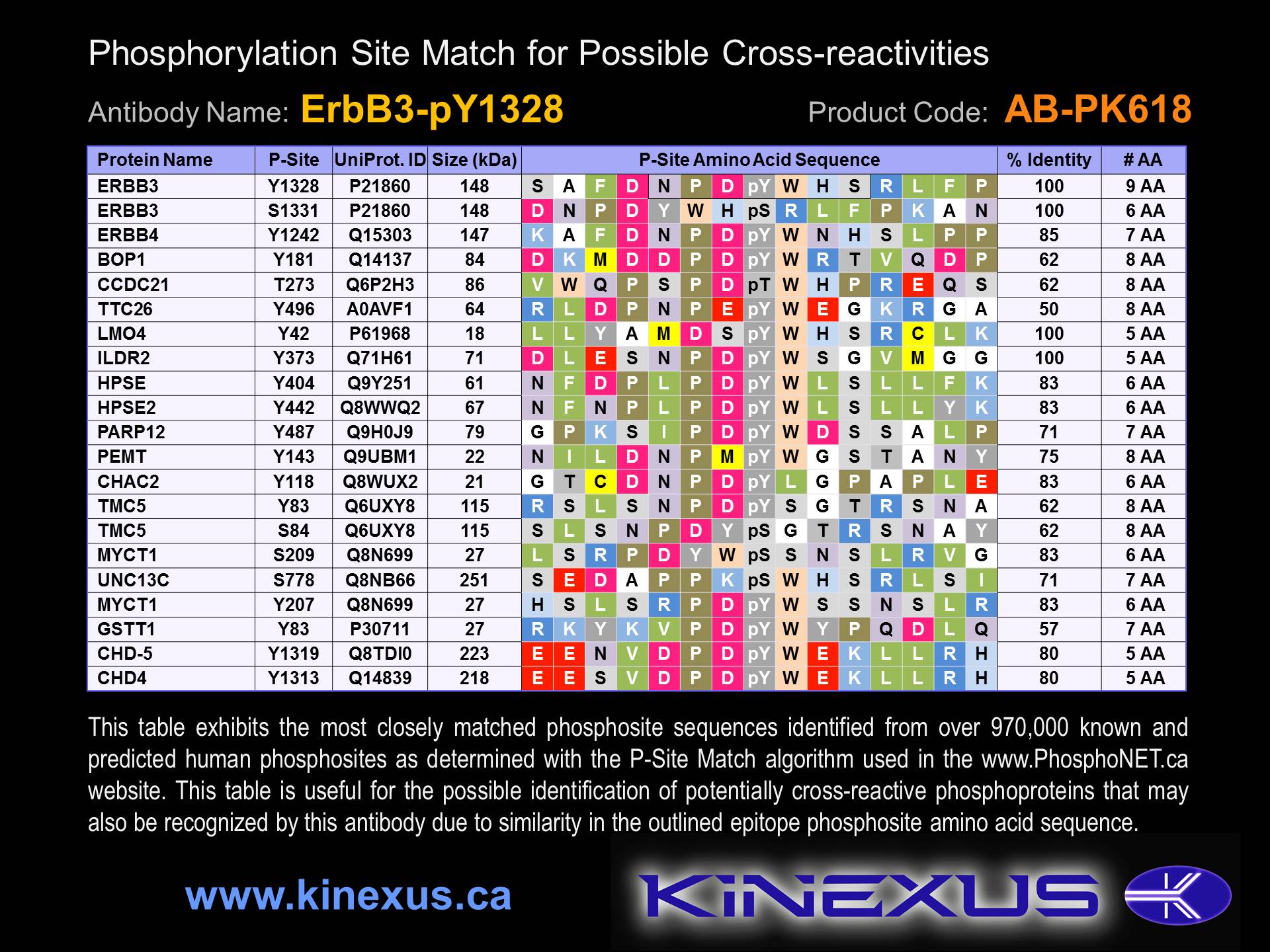
Figure 3. Identification of phosphosites related to ErbB3-pY1328.

© Kinexus Bioinformatics Corporation 2017